. 2005 Dec;6(12):1194-200.
doi: 10.1038/sj.embor.7400540.

Mal3, the fission yeast EB1 homologue, cooperates with Bub1 spindle checkpoint to prevent monopolar attachment

Affiliations

Mal3, the fission yeast EB1 homologue, cooperates with Bub1 spindle checkpoint to prevent monopolar attachment

Kazuhide Asakawa et al. EMBO Rep. 2005 Dec.

Abstract

Bipolar microtubule attachment is central to genome stability. Here, we investigate the mitotic role of the fission yeast EB1 homologue Mal3. Mal3 shows dynamic inward movement along the spindle, initial emergence at the spindle pole body (SPB) and translocation towards the equatorial plane, followed by sudden disappearance. Deletion of Mal3 results in early mitotic delay, which is dependent on the Bub1, but not the Mad2, spindle checkpoint. Consistently, Bub1, but not Mad2, shows prolonged kinetochore localization. Double mutants between mal3 and a subset of checkpoint mutants, including bub1, bub3, mad3 and mph1, but not mad1 or mad2, show massive chromosome mis-segregation defects. In mal3bub1 mutants, both sister centromeres tend to remain in close proximity to one of the separating SPBs. Further analysis indicates that mis-segregated centromeres are exclusively associated with the mother SPB. Mal3, therefore, has a role in preventing monopolar attachment in cooperation with the Bub1/Bub3/Mad3/Mph1-dependent checkpoint.


Figures
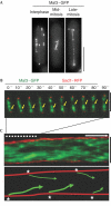
Figure 1
Figure 1
Mitotic localization of Mal3. (A) Mal3–GFP (green fluorescent protein) localization during the cell cycle. (B,C) Time-lapse analysis. Mitotic Mal3–GFP was recorded (B, 10 s intervals) and converted to kymograph (C, upper panel). Inward Mal3 blobs are marked by yellow arrows. White dotted lines in (C) correspond to the first 90 s images shown in (B). In the schematic (C, lower panel, arrows), initial Mal3 loading spots are shown with asterisks. The length of the white arrow represents 1 min. Scale bars, 5 μm (A) or 2 μm (B,C).
Figure 2
Figure 2
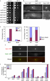
Dependence of mal3 deletion mutants on the Bub1, but not the Mad2, checkpoint. (A) Growth properties of double mutants between mal3 and various spindle checkpoint mutants. WT, wild type. (B) Bub1 localization at the kinetochores. The kinetochore marker (Nuf2–CFP (cyan fluorescent protein)) was used (left panels). As a control, Bub1–GFP (green fluorescent protein) signals, in which Nuf2 was not tagged, were taken using the CFP channel (right panels). (CE) Kinetochore localization of Bub1 and Mad2. Wild type or mal3 mutants that contained Bub1–GFP or Mad2–GFP (C) or Bub1–RFP and Mad2–GFP simultaneously (D) were used for quantification, and representative examples are shown (E, Bub1 in red and Mad2 in green). Scale bar, 3 μm. (F) Chromosome mis-segregation. The percentage of anaphase cells showing chromosome mis-segregation was quantified (n=50).
Figure 3
Figure 3
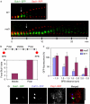
Mitotic localization of Bub1. (A) Time-lapse images. Bub1–GFP (green fluorescent protein) and Sad1–RFP (red fluorescent protein) localization during mitosis in wild type (WT; upper panel) or the mal3 mutant (lower panel) was recorded (n=20) and converted to a kymograph. Arrows indicate the time points when spindle pole body (SPB) separation initiated. (B) Quantification of focal Bub1 signals. The region between the two SPBs was divided into three regions (‘polar' and ‘middle') and localization patterns of Bub1–GFP foci were examined (n=40). (C) Bub1–GFP signals between the two SPBs. The intensity of Bub1–GFP foci just before SPB separation was regarded as 100% (n=8). (D) Close association between Bub1 and Pcp1. Wild-type cells containing Bub1–GFP, Pcp1–RFP and Cut12–CFP (cyan fluorescent protein) were grown at 26°C, starved for nitrogen and resuspended in fresh media to allow re-growth. In the merged image, Bub1–GFP (green), Pcp1–RFP (red) and Cut12–CFP (blue) are shown (n=30). Scale bars, 5 μm (A) or 2 μm (D).
Figure 4
Figure 4
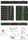
Mono-orientated segregation of sister centromeres in mal3bub1 double mutants. (A) Mis-segregation of sister chromatids. mal3bub1 mutants containing cen2-GFP (green fluorescent protein) were grown at 26°C (left panel, cen2-GFP in green; middle panel, 4,6-diamidino-2-phenylindole (DAPI) in red; right panel, merged). (B,C) Time-lapse analysis. Representative images of wild type (WT; B) and mal3bub1 (C) are shown (cen2-GFP in green and Sad1–RFP (red fluorescent protein) in red). Corresponding kymograph pictures are shown on the right-hand side. The timing of anaphase A onset in wild type and that of anaphase B onset in mal3bub1 cells are marked with a white arrowhead (B) and a double arrowhead (C), respectively. The length of the white arrow represents 1 min. (D) Association of mis-segregated cen2-GFP with the old SPB. Pictures of cen2-GFP (green, left panel), Pcp1–RFP (red, middle panel) and the merged image (right panel) are shown (n=30). Scale bars, 2 μm (B,C, right panels) or 5 μm (B,C (left panels), A,D). (E) Role of Mal3 and the Bub1 spindle checkpoint in sister chromatid segregation. See the text for details.

Similar articles

Cited by

References

    1. Beinhauer JD, Hagan IM, Hegemann JH, Fleig U (1997) Mal3, the fission yeast homologue of the human APC-interacting protein EB-1 is required for microtubule integrity and the maintenance of cell form. J Cell Biol 139: 717–728 - PMC - PubMed
    1. Bernard P, Hardwick K, Javerzat J-P (1998) Fission yeast Bub1 is a mitotic centromere protein essential for the spindle checkpoint and the preservation of correct ploidy through mitosis. J Cell Biol 143: 1775–1787 - PMC - PubMed
    1. Browning H, Hackney DD, Nurse P (2003) Targeted movement of cell end factors in fission yeast. Nat Cell Biol 5: 812–818 - PubMed
    1. Busch KE, Brunner D (2004) The microtubule plus end-tracking proteins mal3p and tip1p cooperate for cell-end targeting of interphase microtubules. Curr Biol 14: 548–559 - PubMed
    1. Cleveland DW, Mao Y, Sullivan KF (2003) Centromeres and kinetochores: from epigenetics to mitotic checkpoint signaling. Cell 112: 407–421 - PubMed

Publication types

MeSH terms