. 2005 Oct;7(10):1021-8.
doi: 10.1038/ncb1302. Epub 2005 Sep 18.

The endoplasmic reticulum gateway to apoptosis by Bcl-X(L) modulation of the InsP3R

Affiliations

The endoplasmic reticulum gateway to apoptosis by Bcl-X(L) modulation of the InsP3R

Carl White et al. Nat Cell Biol. 2005 Oct.

Erratum in

  • Nat Cell Biol. 2006 Mar;8(3):299

Abstract

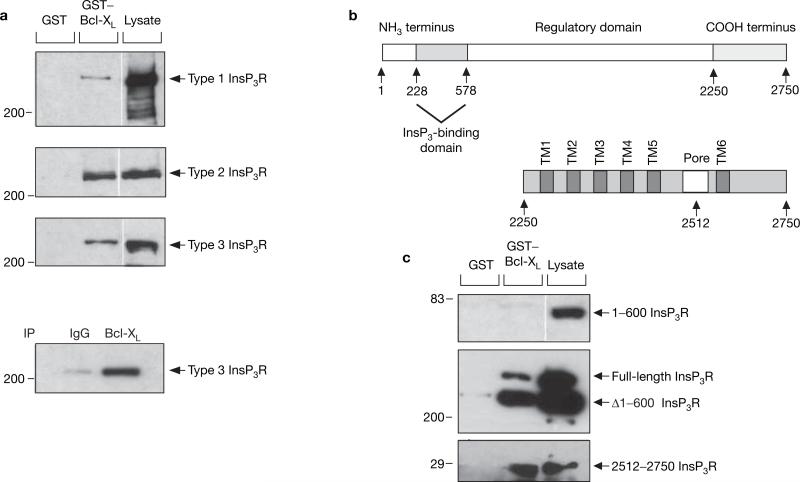
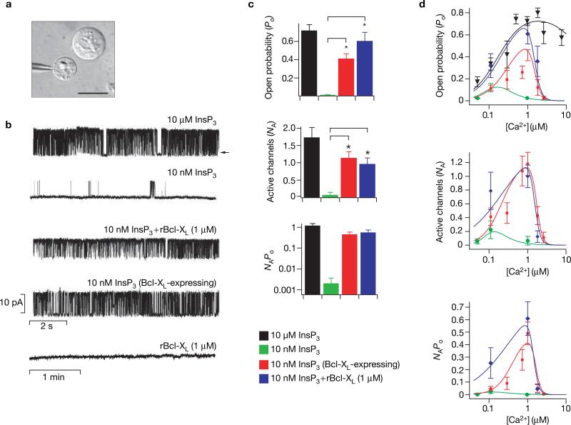
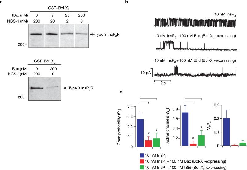
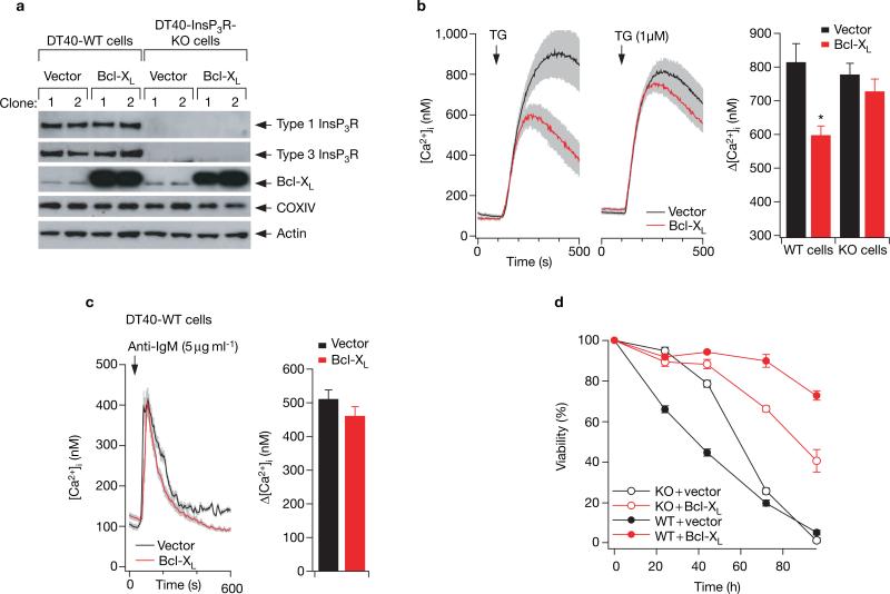
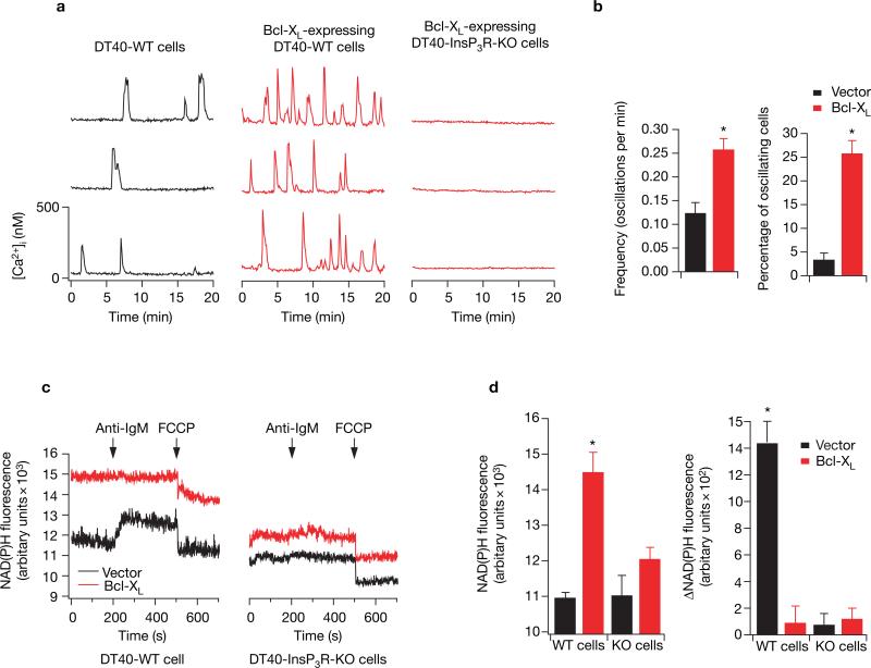
Members of the Bcl-2 protein family modulate outer mitochondrial membrane permeability to control apoptosis. However, these proteins also localize to the endoplasmic reticulum (ER), the functional significance of which is controversial. Here we provide evidence that anti-apoptotic Bcl-2 proteins regulate the inositol 1,4,5-trisphosphate receptor (InsP(3)R) ER Ca(2+) release channel resulting in increased cellular apoptotic resistance and enhanced mitochondrial bioenergetics. Anti-apoptotic Bcl-X(L) interacts with the carboxyl terminus of the InsP(3)R and sensitizes single InsP(3)R channels in ER membranes to low [InsP(3)], enhancing Ca(2+) and InsP(3)-dependent regulation of channel activity in vitro and in vivo, reducing ER Ca(2+) content and stimulating mitochondrial energetics. The pro-apoptotic proteins Bax and tBid antagonize this effect by blocking the biochemical interaction of Bcl-X(L) with the InsP(3)R. These data support a novel model in which Bcl-X(L) is a direct effector of the InsP(3)R, increasing its sensitivity to InsP(3) and enabling ER Ca(2+) release to be more sensitively coupled to extracellular signals. As a consequence, cells are protected against apoptosis by a more sensitive and dynamic coupling of ER to mitochondria through Ca(2+)-dependent signal transduction that enhances cellular bioenergetics and preserves survival.


Figures

Figure 1
Figure 1
Interaction of Bcl-XL with InsP3R. (a) Bcl-XL binds to full-length types 1, 2 and 3 InsP3R. Lysates from DT40-InsP3R-KO cells stably expressing rat type 1 InsP3R and from COS-7 cells that endogenously express type 2 and type 3 InsP3R, were incubated with GST–Bcl-XL, and bound InsP3R was detected with isoform-specific antibodies (top three panels). Bottom panel: co-immunoprecipitation of endogenous Bcl-XL and type 3 InsP3R from COS-7 cells. (b) Domain structure of full-length InsP3R and of its C-terminal region. (c) Bcl-XL binds within the C terminus of InsP3R. GST–Bcl-XL failed to pull-down the V5-tagged 1–600 type 1 InsP3R fragment expressed in COS-7 cells (top panel). Expression of 1–600 InsP3R was verified by a western blot of cell lysates with V5-specific antibody (lane 3). Rat type 1 InsP3R lacking the first 600 residues (Δ1–600 InsP3R) expressed in COS-7 cells was successfully pulled-down along with endogenous InsP3R-1 (middle panel). GST–Bcl-XL effectively binds to the C-terminal 2512–2750 residues of type 1 InsP3R (bottom panel). All western blots depicted are representative of three independent experiments.
Figure 2
Figure 2
Effects of Bcl-XL on InsP3R single-channel activity. (a) Isolated nuclear preparation from Sf9 cells, which endogenously express only type 1 InsP3R. Patch pipette approaching an isolated nucleus with an intact cell visible directly above; scale bar, 15 μM. (b) InsP3R channel activity. Typical InsP3R single-channel current recordings in the presence of saturating (10 μM) or low (10 nM) InsP3; in the absence or presence of recombinant Bcl-XL (rBcl-XL; 1 μM); or with 10 nM InsP3 in cells transiently transfected with Bcl-XL. Channel activity was not evoked by rBcl-XL (1 μM) alone. Pipette [Ca2+] was 1 μM, optimal for channel activity; arrow indicates zero current level. (c) Summary of the effects of Bcl-XL on InsP3R channel activity. In pipettes containing 10 nM InsP3, the open probability (Po) increased from 0.022 ± 0.001 (n = 2) to 0.61 ± 0.09 (n = 10) with addition of 1 μM rBcl-XL (n = number of patches used in Po determination). Similarly, when Bcl-XL was overexpressed, Po increased to 0.42 ± 0.05 (n = 11). Bcl-XL also increased the number of activated channels (NA). In 10 nM InsP3, NA was increased from 0.10 ± 0.07 (n = 20) under control conditions to 1.00 ± 0.16 (n = 54) and 1.18 ± 0.17 (n = 61) in the presence of recombinant or expressed Bcl-XL, respectively. The total ER ion flux as indicated by the product NAPo was increased by Bcl-XL (note log scale). Asterisks indicate P < 0.001, unpaired t-test. Both His- and Flag-tagged rBcl-XL, generated using distinct purification protocols, were equally effective, whereas His-tagged NCS-1, which does not interact with InsP3R (ref. 32), had no effect either alone or in combination with 10 nM InsP3 (data not shown). (d) Dependence of InsP3R channel activity on [Ca2+] at the cytoplasmic face of the channel. Effect of [Ca2+]i on Po, NA and NAPo in the presence of 10 μM InsP3 (black inverted triangles), 10 nM InsP3 (green circles), 10 nM InsP3 + 1 μM rBcl-XL (blue diamonds) and 10 nM InsP3 + Bcl-XL expression (red squares).
Figure 3
Figure 3
tBid and Bax antagonize the effects of Bcl-XL on InsP3R channel activity. (a) tBid and Bax inhibit binding of Bcl-XL to InsP3R. COS-7 cell lysates were incubated with GST–Bcl-XL in the absence or presence of recombinant tBid (2–200 nM; upper panel) or recombinant Bax (200 nM; lower panel), and bound InsP3R was detected with a type 3 antibody. Recombinant neuronal calcium sensor-1 (NCS-1) was used to control for total protein concentration in both experiments. Data are representative of three independent experiments. (b) tBid and Bax inhibit the electrophysiological effects of Bcl-XL. Typical current traces showing the effects of 100 nM recombinant tBid or Bax in the presence of 10 nM InsP3 in nuclei isolated from cells transiently transfected with Bcl-XL. Pipette [Ca2+] was 1 μM. (c) Addition of 100 nM tBid or Bax decreased Po from 0.28 ± 0.06 (n = 5) to 0.07 ± 0.03 (n = 3) and 0.09 ± 0.05 (n = 3), respectively. Similarly, NA was reduced from 0.74 ± 0.14 (n = 38) to 0.26 ± 0.10 (n = 33) in the presence of tBid and to 0.09 ± 0.05 (n = 33) in the presence of Bax. The product NAPo was also reduced. Asterisks indicate P < 0.001, unpaired t-test.
Figure 4
Figure 4
Interaction of Bcl-XL with InsP3R is essential for Bcl-XL effects on ER Ca2+ regulation and inhibition of apoptosis. (a) The empty vectors pIRES2-DsRed2 or pBcl-XL-IRES2-DsRed2 were stably expressed in DT40-WT and DT40-InsP3R-KO cells. Expression levels of types 1 and 3 InsP3R, Bcl-XL and OxPhos complex IV (COXIV; subunit 1) were examined by western blot. Expression of the mitochondrial complex IV protein was unchanged in the Bcl-XL-expressing clones. Depicted blots are representative of three independent experiments. (b) Effects of Bcl-XL expression on the Ca2+ content of the ER (Ca2+ER). Typical records depicting change in cytoplasmic [Ca2+] ([Ca2+]i) in response to application of 1 μM thapsigargin (TG) in DT40-WT and DT40-InsP3R-KO cells stably transfected with either Bcl-XL or vector alone. Ca2+ER was indirectly estimated by single-cell imaging of the [Ca2+]i responses to acute inhibition by thapsigargin of ER Ca2+ uptake. Each trace represents mean ± s.e.m. of at least six cells within the image field. Bar graph summarizes the effects of thapsigargin; data represent mean ± s.e.m. for at least 30 cells in multiple trials. Asterisk indicates P < 0.05, ANOVA. Mean values of resting [Ca2+]i ranged from 80 to 100 nM, with no significant differences among duplicate clones of the four cell lines (data not shown). (c) [Ca2+]i transients in response to 5 μg ml−1 anti-BCR antibody (anti-IgM) in DT40-WT cells stably transfected with either Bcl-XL or vector alone. Summary data represent the peak amplitude (mean ± s.e.m.) for at least 30 cells in multiple trials. (d) Cell viability after treatment with 20 μg ml−1 anti-BCR antibody (anti-IgM) (time 0) of DT40-WT (solid symbols) and DT40-InsP3R-KO (open symbols) cells stably expressing Bcl-XL (red) or vector alone (same clones as in b). Similar results were obtained using independent clones (see Supplementary Information, Fig. S3).
Figure 5
Figure 5
The Bcl-XL–InsP3R interaction modulates [Ca2+]i signalling and mitochondrial NADH levels. (a) Spontaneous [Ca2+]i oscillations in three representative DT40-WT cells stably expressing vector alone (left) or Bcl-XL (middle), and DT40-InsP3R-KO cells expressing Bcl-XL (right).(b) The difference in frequency and number of oscillating cells (mean ± s.e.m.) between vector only and Bcl-XL-expressing cells. (c) NAD(P)H fluorescence measurements from DT40-WT and DT40-InsP3R-KO cells expressing Bcl-XL or vector in response to 5 μg ml−1 anti-BCR antibody (anti-IgM) and FCCP (2 μM). The average (± s. e.m.) resting NAD(P)H fluorescence and the change in NAD(P)H fluorescence in response to anti-IgM stimulation for four independent experiments are plotted in d. Asterisk indicates P < 0.001, ANOVA. Similar results were obtained using independent clones (see Supplementary Information, Fig. S4).

Similar articles

Cited by

References

    1. Wei MC, et al. Proapoptotic BAX and BAK: A requisite gateway to mitochondrial dysfunction and death. Science. 2001;292:727–730. - PMC - PubMed
    1. Vander Heiden MG, Thompson CB. Bcl-2 proteins: regulators of apoptosis or of mitochondrial homeostasis? Nature Cell Biol. 1999;1:E209–E216. - PubMed
    1. Krajewski S, et al. Investigation of the subcellular distribution of the Bcl-2 oncoprotein - residence in the nuclear envelope, endoplasmic reticulum, and outer mitochondrial membranes. Cancer Res. 1993;53:4701–4714. - PubMed
    1. Distelhorst CW, Shore GC. Bcl-2 and calcium: controversy beneath the surface. Oncogene. 2004;23:2875–2880. - PubMed
    1. Jurgensmeier JM, et al. Bax directly induces release of cytochrome c from isolated mitochondria. Proc. Natl Acad. Sci. USA. 1998;95:4997–5002. - PMC - PubMed

Publication types

MeSH terms