

Impaired response to amphetamine and neuronal degeneration in the nucleus accumbens of autoimmune MRL-lpr mice
- PMID: 16183144
- PMCID: PMC1634760
- DOI: 10.1016/j.bbr.2005.07.030
Impaired response to amphetamine and neuronal degeneration in the nucleus accumbens of autoimmune MRL-lpr mice
Abstract
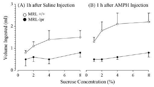
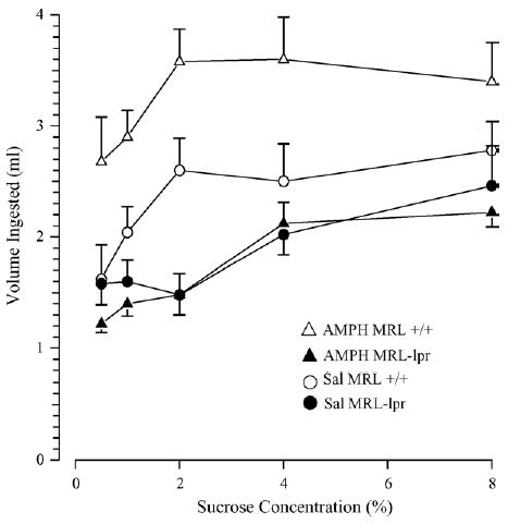
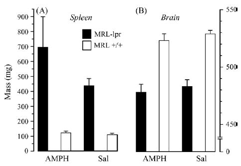
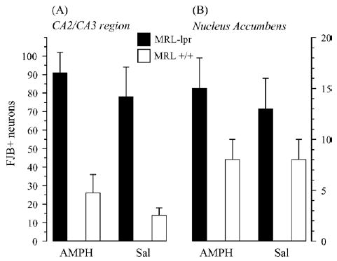
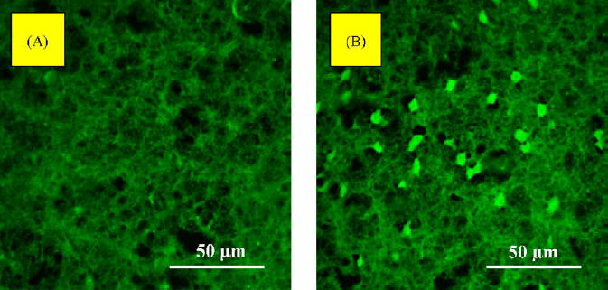
Spontaneous development of lupus-like disease in MRL-lpr mice is accompanied by a constellation of behavioral deficits, including blunted responsiveness to sucrose. Although autoimmunity-induced damage of limbic areas is proposed to underlie this deficit, the systemic nature of the disease precludes inference of a causal relationship between CNS damage and functional loss. Based on the stimulatory effects of d-amphetamine sulfate (AMPH) on sucrose intake, the present study pharmacologically probes the functional status of central dopaminergic circuits involved in control of behavioral reward. The response rates were compared between diseased MRL-lpr mice and congenic MRL +/+ controls tested in the sucrose preference paradigm. Neuronal loss was assessed by Fluoro Jade B (FJB) staining of nucleus accumbens and the CA2/CA3 region. While control mice significantly increased intake of sucrose solutions 60 min after administration of AMPH (i.p., 0.5 mg/kg), the intake in drugged MRL-lpr mice was comparable to those given saline injection. Increased FJB staining was detected in the nucleus accumbens and hippocampus of diseased mice, and AMPH treatment neither altered this nor other measures of organ pathology. The results obtained are consistent with previously observed changes in the mesolimbic dopamine system of MRL-lpr mice and suggest that the lesion in the nucleus accumbens and deficits in dopamine release underlie impaired responsiveness to palatable stimulation during the progress of systemic autoimmune disease. As such, they point to a neurotransmitter-specific regional brain damage which may account for depressive behaviors in neuropsychiatric lupus erythematosus.
Figures
Similar articles
-
Autoimmune-induced damage of the midbrain dopaminergic system in lupus-prone mice.J Neuroimmunol. 2004 Jul;152(1-2):83-97. doi: 10.1016/j.jneuroim.2004.04.003. J Neuroimmunol. 2004. PMID: 15223241
-
Hippocampal damage in mouse and human forms of systemic autoimmune disease.Hippocampus. 2004;14(5):649-61. doi: 10.1002/hipo.10205. Hippocampus. 2004. PMID: 15301441 Free PMC article.
-
Neurodegeneration in autoimmune MRL-lpr mice as revealed by Fluoro Jade B staining.Brain Res. 2003 Feb 28;964(2):200-10. doi: 10.1016/s0006-8993(02)03980-x. Brain Res. 2003. PMID: 12576180
-
The MRL Model: A Valuable Tool in Studies of Autoimmunity-Brain Interactions.Methods Mol Biol. 2018;1781:259-285. doi: 10.1007/978-1-4939-7828-1_14. Methods Mol Biol. 2018. PMID: 29705852 Review.
-
The MRL Model: A Valuable Tool in Studies of Autoimmunity-Brain Interactions.Methods Mol Biol. 2025;2868:221-246. doi: 10.1007/978-1-0716-4200-9_12. Methods Mol Biol. 2025. PMID: 39546233 Review.
Cited by
-
Effects of prolonged treatment with memantine in the MRL model of CNS lupus.Clin Exp Neuroimmunol. 2012 Sep;3(3):116-128. doi: 10.1111/j.1759-1961.2012.00032.x. Clin Exp Neuroimmunol. 2012. PMID: 23554849 Free PMC article.
-
Impaired decision-making and functional neuronal network activity in systemic lupus erythematosus.J Magn Reson Imaging. 2018 Dec;48(6):1508-1517. doi: 10.1002/jmri.26006. Epub 2018 Mar 14. J Magn Reson Imaging. 2018. PMID: 29537670 Free PMC article.
-
The role of tumor necrosis factor receptor superfamily members in mammalian brain development, function and homeostasis.Rev Neurosci. 2011;22(5):509-33. doi: 10.1515/RNS.2011.041. Epub 2011 Aug 24. Rev Neurosci. 2011. PMID: 21861782 Free PMC article. Review.
-
Ibuprofen fails to prevent brain pathology in a model of neuropsychiatric lupus.J Rheumatol. 2006 Nov;33(11):2199-213. J Rheumatol. 2006. PMID: 17086606 Free PMC article.
-
Fluoro-Jade B staining following zymosan microinjection into the spinal cord white matter.Cell Mol Neurobiol. 2006 Oct-Nov;26(7-8):1463-73. doi: 10.1007/s10571-006-9081-5. Epub 2006 Jun 14. Cell Mol Neurobiol. 2006. PMID: 16773443 Free PMC article.
References
-
- Allan SM, Rothwell NJ. Cytokines and acute neurodegeneration. Nat Rev Neurosci. 2001;2:734–44. - PubMed
-
- Anisman H, Merali Z. Cytokines, stress and depressive illness: brain–immune interactions. Ann Med. 2003;35:2–11. - PubMed
-
- Anisman H, Merali Z, Poulter MO, Hayley S. Cytokines as a precipitant of depressive illness: animal and human studies. Curr Pharm Des. 2005;11:963–72. - PubMed
-
- Ballok DA, Earls AM, Krasnik C, Hoffman SA, Sakic B. Autoimmune-induced damage of the midbrain dopaminergic system in lupus-prone mice. J Neuroimmunol. 2004;152:83–97. - PubMed
-
- Ballok DA, Millward JM, Sakic B. Neurodegeneration in autoimmune MRL-lpr mice as revealed by Fluoro Jade B staining. Brain Res. 2003;964:200–10. - PubMed
Publication types
MeSH terms
Substances
Grants and funding
LinkOut - more resources
Full Text Sources
Medical
Research Materials
Miscellaneous
