Cysteine protease mcII-Pa executes programmed cell death during plant embryogenesis
- PMID: 16183741
- PMCID: PMC1242326
- DOI: 10.1073/pnas.0506948102
Cysteine protease mcII-Pa executes programmed cell death during plant embryogenesis
Abstract
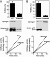
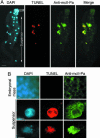
Programmed cell death (PCD) is indispensable for eukaryotic development. In animals, PCD is executed by the caspase family of cysteine proteases. Plants do not have close homologues of caspases but possess a phylogenetically distant family of cysteine proteases named metacaspases. The cellular function of metacaspases in PCD is unknown. Here we show that during plant embryogenesis, metacaspase mcII-Pa translocates from the cytoplasm to nuclei in terminally differentiated cells that are destined for elimination, where it colocalizes with the nuclear pore complex and chromatin, causing nuclear envelope disassembly and DNA fragmentation. The cell-death function of mcII-Pa relies on its cysteine-dependent arginine-specific proteolytic activity. Accordingly, mutation of catalytic cysteine abrogates the proteolytic activity of mcII-Pa and blocks nuclear degradation. These results establish metacaspase as an executioner of PCD during embryo patterning and provide a functional link between PCD and embryogenesis in plants. Although mcII-Pa and metazoan caspases have different substrate specificity, they serve a common function during development, demonstrating the evolutionary parallelism of PCD pathways in plants and animals.
Figures


Similar articles
-
Tudor staphylococcal nuclease is an evolutionarily conserved component of the programmed cell death degradome.Nat Cell Biol. 2009 Nov;11(11):1347-54. doi: 10.1038/ncb1979. Epub 2009 Oct 11. Nat Cell Biol. 2009. PMID: 19820703
-
Plant life needs cell death, but does plant cell death need Cys proteases?FEBS J. 2017 May;284(10):1577-1585. doi: 10.1111/febs.14034. Epub 2017 Feb 26. FEBS J. 2017. PMID: 28165668 Review.
-
Metacaspase-8 modulates programmed cell death induced by ultraviolet light and H2O2 in Arabidopsis.J Biol Chem. 2008 Jan 11;283(2):774-83. doi: 10.1074/jbc.M704185200. Epub 2007 Nov 12. J Biol Chem. 2008. PMID: 17998208
-
Metacaspase-dependent programmed cell death is essential for plant embryogenesis.Curr Biol. 2004 May 4;14(9):R339-40. doi: 10.1016/j.cub.2004.04.019. Curr Biol. 2004. PMID: 15120084 No abstract available.
-
Caspases in plants: metacaspase gene family in plant stress responses.Funct Integr Genomics. 2015 Nov;15(6):639-49. doi: 10.1007/s10142-015-0459-7. Epub 2015 Aug 16. Funct Integr Genomics. 2015. PMID: 26277721 Review.
Cited by
-
Proteome profiling of early seed development in Cunninghamia lanceolata (Lamb.) Hook.J Exp Bot. 2010 May;61(9):2367-81. doi: 10.1093/jxb/erq066. Epub 2010 Apr 2. J Exp Bot. 2010. PMID: 20363864 Free PMC article.
-
Protein S-nitrosylation in programmed cell death in plants.Cell Mol Life Sci. 2019 May;76(10):1877-1887. doi: 10.1007/s00018-019-03045-0. Epub 2019 Feb 19. Cell Mol Life Sci. 2019. PMID: 30783684 Free PMC article. Review.
-
Calcium-dependent activation and autolysis of Arabidopsis metacaspase 2d.J Biol Chem. 2011 Mar 25;286(12):10027-40. doi: 10.1074/jbc.M110.194340. Epub 2011 Jan 5. J Biol Chem. 2011. PMID: 21209078 Free PMC article.
-
Indispensable Role of Proteases in Plant Innate Immunity.Int J Mol Sci. 2018 Feb 23;19(2):629. doi: 10.3390/ijms19020629. Int J Mol Sci. 2018. PMID: 29473858 Free PMC article. Review.
-
Biochemical and Bioinformatic Characterization of Type II Metacaspase Protein (TaeMCAII) from Wheat.Plant Mol Biol Report. 2012;30(6):1338-1347. doi: 10.1007/s11105-012-0450-6. Plant Mol Biol Report. 2012. PMID: 24415839 Free PMC article.
References
-
- Baehrecke, E. (2002) Nat. Rev. Mol. Cell Biol. 3, 779-787. - PubMed
-
- Bozhkov, P. V., Suarez, M. F. & Filonova, L. H. (2005) Curr. Top. Dev. Biol. 67, 135-179. - PubMed
-
- Goldberg, R. B., de Paiva, G. & Yadegari, R. (1994) Science 266, 605-614. - PubMed
-
- Lukowitz, W., Roeder, A., Parmenter, D. & Somerville, C. (2004) Cell 116, 109-119. - PubMed
-
- Earnshaw, W. C. (1995) Curr. Opin. Cell Biol. 7, 337-343. - PubMed
Publication types
MeSH terms
Substances
Associated data
- Actions
LinkOut - more resources
Full Text Sources

