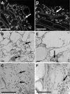
Pseudomonas fluorescens and Glomus mosseae trigger DMI3-dependent activation of genes related to a signal transduction pathway in roots of Medicago truncatula
- PMID: 16183836
- PMCID: PMC1256018
- DOI: 10.1104/pp.105.067603
Pseudomonas fluorescens and Glomus mosseae trigger DMI3-dependent activation of genes related to a signal transduction pathway in roots of Medicago truncatula
Abstract
Plant genes induced during early root colonization of Medicago truncatula Gaertn. J5 by a growth-promoting strain of Pseudomonas fluorescens (C7R12) have been identified by suppressive subtractive hybridization. Ten M. truncatula genes, coding proteins associated with a putative signal transduction pathway, showed an early and transient activation during initial interactions between M. truncatula and P. fluorescens, up to 8 d after root inoculation. Gene expression was not significantly enhanced, except for one gene, in P. fluorescens-inoculated roots of a Myc(-)Nod(-) genotype (TRV25) of M. truncatula mutated for the DMI3 (syn. MtSYM13) gene. This gene codes a Ca(2+) and calmodulin-dependent protein kinase, indicating a possible role of calcium in the cellular interactions between M. truncatula and P. fluorescens. When expression of the 10 plant genes was compared in early stages of root colonization by mycorrhizal and rhizobial microsymbionts, Glomus mosseae activated all 10 genes, whereas Sinorhizobium meliloti only activated one and inhibited four others. None of the genes responded to inoculation by either microsymbiont in roots of the TRV25 mutant. The similar response of the M. truncatula genes to P. fluorescens and G. mosseae points to common molecular pathways in the perception of the microbial signals by plant roots.
Figures


Similar articles
-
Fungal elicitation of signal transduction-related plant genes precedes mycorrhiza establishment and requires the dmi3 gene in Medicago truncatula.Mol Plant Microbe Interact. 2004 Dec;17(12):1385-93. doi: 10.1094/MPMI.2004.17.12.1385. Mol Plant Microbe Interact. 2004. PMID: 15597744
-
Common gene expression in Medicago truncatula roots in response to Pseudomonas fluorescens colonization, mycorrhiza development and nodulation.New Phytol. 2004 Mar;161(3):855-863. doi: 10.1046/j.1469-8137.2004.00997.x. Epub 2004 Jan 8. New Phytol. 2004. PMID: 33873727
-
Identification of new potential regulators of the Medicago truncatula-Sinorhizobium meliloti symbiosis using a large-scale suppression subtractive hybridization approach.Mol Plant Microbe Interact. 2007 Mar;20(3):321-32. doi: 10.1094/MPMI-20-3-0321. Mol Plant Microbe Interact. 2007. PMID: 17378435
-
Combined genetic and transcriptomic analysis reveals three major signalling pathways activated by Myc-LCOs in Medicago truncatula.New Phytol. 2015 Oct;208(1):224-40. doi: 10.1111/nph.13427. Epub 2015 Apr 28. New Phytol. 2015. PMID: 25919491
-
Annexins - calcium- and membrane-binding proteins in the plant kingdom: potential role in nodulation and mycorrhization in Medicago truncatula.Acta Biochim Pol. 2009;56(2):199-210. Epub 2009 May 7. Acta Biochim Pol. 2009. PMID: 19421430 Review.
Cited by
-
Fungal and plant gene expression in arbuscular mycorrhizal symbiosis.Mycorrhiza. 2006 Nov;16(8):509-524. doi: 10.1007/s00572-006-0069-2. Epub 2006 Sep 27. Mycorrhiza. 2006. PMID: 17004063 Review.
-
Two Medicago truncatula growth-promoting rhizobacteria capable of limiting in vitro growth of the Fusarium soil-borne pathogens modulate defense genes expression.Planta. 2023 May 12;257(6):118. doi: 10.1007/s00425-023-04145-9. Planta. 2023. PMID: 37173556 Free PMC article.
-
Unique and common traits in mycorrhizal symbioses.Nat Rev Microbiol. 2020 Nov;18(11):649-660. doi: 10.1038/s41579-020-0402-3. Epub 2020 Jul 21. Nat Rev Microbiol. 2020. PMID: 32694620 Review.
-
Bioinformatic and empirical analysis of a gene encoding serine/threonine protein kinase regulated in response to chemical and biological fertilizers in two maize (Zea mays L.) cultivars.Mol Biol Res Commun. 2017 Jun;6(2):65-75. Mol Biol Res Commun. 2017. PMID: 28775992 Free PMC article.
-
Alfalfa snakin-1 prevents fungal colonization and probably coevolved with rhizobia.BMC Plant Biol. 2014 Sep 17;14:248. doi: 10.1186/s12870-014-0248-9. BMC Plant Biol. 2014. PMID: 25227589 Free PMC article.
References
-
- Allende JE, Allende CC (1995) Protein kinase CK2: an enzyme with multiple substrates and puzzling regulation. FASEB J 9: 313–323 - PubMed
-
- Ané JM, Kiss GB, Riely BK, Pensmetsa RV, Oldroyd GED, Ayax C, Lévy J, Debellé F, Baek JM, Kalo P, et al (2004) Medicago truncatula DMI1 required for bacterial and fungal symbioses in legumes. Science 303: 1364–1367 - PubMed
Publication types
MeSH terms
Substances
Associated data
- Actions
- Actions
- Actions
- Actions
- Actions
- Actions
- Actions
- Actions
- Actions
- Actions
- Actions
- Actions
- Actions
- Actions
- Actions
- Actions
- Actions
- Actions
- Actions
- Actions
- Actions
- Actions
- Actions
- Actions
- Actions
- Actions
- Actions
- Actions
- Actions
- Actions
- Actions
- Actions
- Actions
- Actions
- Actions
- Actions
- Actions
- Actions
- Actions
- Actions
- Actions
- Actions
- Actions
- Actions
- Actions
- Actions
- Actions
- Actions
- Actions
- Actions
- Actions
- Actions
- Actions
- Actions
- Actions
- Actions
- Actions
- Actions
LinkOut - more resources
Full Text Sources
Medical
Miscellaneous

