. 2005 Sep;171(1):35-47.
doi: 10.1534/genetics.105.040634.

Dissection of Arp2/3 complex actin nucleation mechanism and distinct roles for its nucleation-promoting factors in Saccharomyces cerevisiae

Affiliations

Dissection of Arp2/3 complex actin nucleation mechanism and distinct roles for its nucleation-promoting factors in Saccharomyces cerevisiae

Jessica L D'Agostino et al. Genetics. 2005 Sep.

Abstract

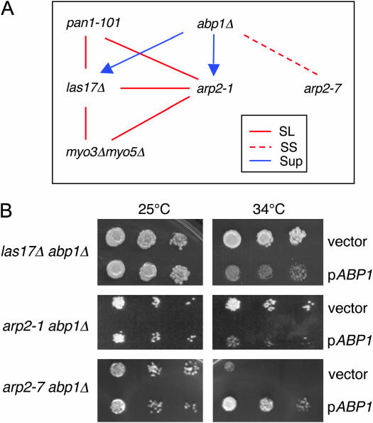
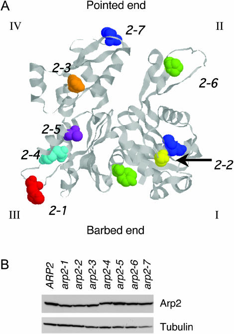
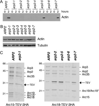
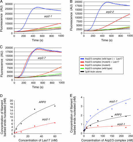
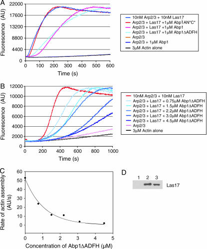
Actin nucleation by the Arp2/3 complex is under tight control, remaining inactive until stimulation by nucleation-promoting factors (NPFs). Although multiple NPFs are expressed in most cell types, little is known about how they are coordinated and whether they perform similar or distinct functions. We examined genetic relationships among the four S. cerevisiae NPFs. Combining las17delta with pan1-101 or myo3delta myo5delta was lethal at all temperatures, whereas combining pan1-101 with myo3delta myo5delta showed no genetic interaction and abp1delta partially suppressed las17delta. These data suggest that NPFs have distinct and overlapping functions in vivo. We also tested genetic interactions between each NPF mutant and seven different temperature-sensitive arp2 alleles and purified mutant Arp2/3 complexes to compare their activities. Two arp2 alleles with mutations at the barbed end were severely impaired in nucleation, providing the first experimental evidence that Arp2 nucleates actin at its barbed end in vitro and in vivo. Another arp2 allele caused partially unregulated ("leaky") nucleation in the absence of NPFs. Combining this mutant with a partially unregulated allele in a different subunit of Arp2/3 complex was lethal, suggesting that cells cannot tolerate high levels of unregulated activity. Genetic interactions between arp2 alleles and NPF mutants point to Abp1 having an antagonistic role with respect to other NPFs, possibly serving to attenuate their stronger activities. In support of this model, Abp1 binds strongly to Arp2/3 complex, yet has notably weak nucleation-promoting activity and inhibits Las17 activity on Arp2/3 complex in a dose-responsive manner.


Figures

Figure 1.
Figure 1.
Genetic interactions among nucleation-promoting factor mutants and arp2 alleles. (A) Diagram of genetic relationships. Six haploid strains bearing las17Δ, abp1Δ, myo3Δ myo5Δ, pan1-101, arp2-1, and arp2-7 mutations were crossed in all pairwise combinations to test genetic interactions. The data for arp2 alleles are summarized in Table 2. The absence of a line indicates that no interaction was detected. SL, synthetic lethal; SS, synthetic sick; SUP, suppression. The direction of the arrow indicates direction of suppression. Thus, the growth defects of both arp2-1 and las17Δ strains are partially suppressed by abp1Δ. (B) Differential genetic interactions of abp1Δ with las17Δ, arp2-1, and arp2-7 mutations. Haploid abpΔ las17Δ, abp1Δ arp2-1, and abp1Δ arp2-7 strains were transformed with a pABP1 CEN expression plasmid (Lila and Drubin 1997) or empty vector, plated on selective media, and grown at 25° and 34°.
Figure 2.
Figure 2.
Positions of residues mutated in arp2 alleles and Arp2 expression levels in the mutant strains. (A) The residues mutated in each arp2 allele (Table 1) are highlighted on the crystal structure of rabbit skeletal muscle actin (1ATN). The crystal structure of Arp2 is not shown because only half of its structure is solved (Robinson et al. 2001). The four subdomains (I, II, III, and IV) are indicated, and the Arp2-specific loop in subdomain III was inserted from a crystallized portion of Arp2 (1K8K). The specific residues mutated in each allele are shown in space-fill and color-coded: arp2-1 (red), arp2-2 (yellow), arp2-3 (orange), arp2-4 (cyan), arp2-5 (purple), arp2-6 (green), and arp2-7 (blue). (B) Arp2 protein levels were compared in wild-type and arp2 mutant cells grown to log phase by immunoblotting total cell extracts with Arp2 antibodies. The same samples were immunoblotted with tubulin antibodies as a loading control. Although some lanes on the tubulin blot show a lower signal (e.g., arp2-7), repeated experiments demonstrated that variation was due to inefficient transfer of some lanes and that tubulin and Arp2 levels are similar in whole-cell extracts from these strains (not shown).
Figure 3.
Figure 3.
Purification of Arp2/3 complex from wild-type and arp2 mutant strains. (A) Clarified cell extracts isolated from wild-type and arp2 strains were compared for their ability to nucleate actin assembly over time. Samples of reactions were removed at time zero and after 2 hr of incubation at 4° and centrifuged at 80,000 × g to pellet polymerized actin (Goode 2002). Actin levels in the pellets were analyzed by SDS-PAGE and immunoblotting with actin antibodies; >50% of the cellular actin assembles in wild-type extracts (A. Goodman, J. L. D'Agostino and B. L. Goode, unpublished results). (B) Starting actin levels in the clarified extracts were compared by immunoblotting with actin antibodies. The same samples were immunoblotted with tubulin antibodies as a loading control. (C) Coomassie-stained gels of Arp2/3 complex isolated from wild-type (ARP2) and arp2 strains. A TEV-3xHA affinity tag integrated at the C terminus of the Arc15/ARPC5 subunit was used to isolate Arp2/3 complex from wild-type and arp2-1 strains. A similar tag on the Arc18/ARPC3 subunit was used to isolate Arp2/3 complex from wild-type, arp2-2, arp2-5, arp2-6, and arp2-7 strains. A remnant of nine residues is left on the indicated subunit after TEV protease digestion, causing the aberrant migration of that subunit (*).
Figure 4.
Figure 4.
Actin nucleation activities of Arp2/3 complexes isolated from wild-type and mutant arp2 strains. (A–C) Assembly of monomeric actin (3 μm, 5% pyrene labeled) was compared in the presence and absence of 15 nm full-length Las17 and 15 nm wild type or arp2-1 (A), arp2-2 (B) or arp2-7 (C) Arp2/3 complex. (D) Comparison of the actin nucleation activities of wild-type and arp2-1 Arp2/3 complexes (15 nm) in the presence of variable concentrations of Las17. (E) Quantitative comparison of actin nucleation activities for wild-type, arp2-1, and arp2-7 Arp2/3 complexes in the absence of Las17.
Figure 5.
Figure 5.
Purified Abp1 attenuates the nucleation-promoting activity of Las17 on Arp2/3 complex in a dose-responsive manner. (A) Monomeric actin (3 μm, 5% pyrene-labeled) was polymerized in the presence of Arp2/3 complex (15 nm), Las17 (15 nm), and 1μm of each of the following proteins: full-length wild-type Abp1, Abp1ΔADFH, and Abp1-AN*C* (mutated to disrupt both of its acidic motifs). (B) Monomeric actin (as above) was polymerized in the presence of Arp2/3 complex (15 nm), Las17 (15 nm), and a range of concentrations of Abp1ΔADFH. (C) Graph showing the concentration-dependent effects of Abp1ΔADFH on rate of actin assembly induced by Las17 and Arp2/3 complex. (D) Association of Las17 (30 nm) with Arp2/3 complex (15 nm) is partially disrupted in the presence of excess Abp1 (2 μm). Beads coated with Arc18-HA-tagged Arp2/3 complex or control beads were incubated for 10 min with Las17 in the presence or absence of Abp1. Bead pellets were immunoblotted with anti-VCA antibody: Lane 1, control beads; lane 2, Arp2/3 beads without Abp1; lane 3, Arp2/3 beads with Abp1.

References

    1. Beltzner, C. C., and T. D. Pollard, 2004. Identification of functionally important residues of Arp2/3 complex by analysis of homology models from diverse species. J. Mol. Biol. 336: 551–565. - PubMed
    1. Duncan, M. C., M. J. Cope, B. L. Goode, B. Wendland and D. G. Drubin, 2001. Yeast Eps15-like endocytic protein, Pan1p, activates the Arp2/3 complex. Nat. Cell Biol. 3: 687–690. - PubMed
    1. Evangelista, M., B. M. Klebl, A. H. Tong, B. A. Webb, T. Leeuw et al., 2000. A role for myosin-I in actin assembly through interactions with Vrp1p, Bee1p, and the Arp2/3 complex. J. Cell Biol. 148: 353–362. - PMC - PubMed
    1. Goley, E. D., S. E. Rodenbusch, A. C. Martin and M. D. Welch, 2004. Critical conformational changes in the Arp2/3 complex are induced by nucleotide and nucleation promoting factor. Mol. Cell 16: 269–279. - PubMed
    1. Goode, B. L., 2002. Purification of yeast actin and actin-associated proteins. Methods Enzymol. 351: 433–441. - PubMed

Publication types

MeSH terms