

Review
doi: 10.1038/nrd1853.
Viral and cellular RNA helicases as antiviral targets
Affiliations
- PMID: 16184083
- PMCID: PMC7097191
- DOI: 10.1038/nrd1853
Item in Clipboard
Review
Viral and cellular RNA helicases as antiviral targets
Nat Rev Drug Discov.
2005 Oct.
Abstract
Although there has been considerable progress in the development of antiviral agents in recent years, there is still a pressing need for new drugs both to improve on the properties of existing agents and to combat the problem of viral resistance. Helicases, both viral and human, have recently emerged as novel targets for the treatment of viral infections. Here, we discuss the role of these enzymes, factors affecting their potential as drug targets and progress in the development of agents that inhibit their activity using the hepatitis C virus-encoded helicase NS3 and the cellular helicase DDX3 adopted for use by HIV-1 as examples.
Conflict of interest statement
The authors declare no competing financial interests.
Figures
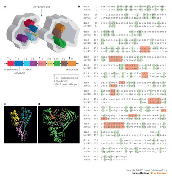
a | Schematic illustration of a two-domain SF2 helicase with the consensus motifs as indicated. b | Sequence alignment of the hepatitis C virus (HCV) NS3 helicase and the DDX3 cellular helicase used by HIV-1 as a Rev co-factor. Analogous motifs between the two SF2 helicases are highlighted. Note the paucity of sequence relatedness outside of the helicase motifs. c | The three-domain structure of HCV NS3 helicase with the bound poly(U) (PDB code: 1A1V). Domains 1, 2 and 3 are coloured in magenta, yellow and cyan, respectively. d | The poly(U), coloured orange, binds at the interface of domain 3 with the first two domains. Illustration of position of consensus motifs (highlighted in orange) on the inner faces of domains 1 and 2 using HCV helicase structure. The position of an oligonucleotide from a co-complex structure is shown; however, this is not conserved in other helicases.
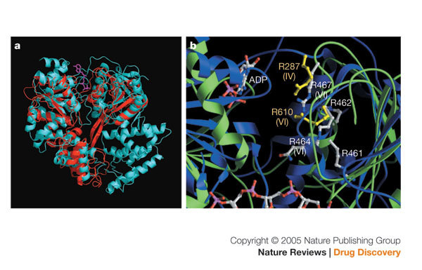
a | The overlay of the crystal structures of hepatitis C virus (HCV) helicase (red; PDB code: 1HEI) and PcrA helicase (cyan; PDB code: 1QHH ). The ATP is coloured magenta. The HCV helicase has three domains, whereas the PcrA helicase has four domains. The overlay shows the overall structural homology of three domains of the two structures, although they share very little sequence similarity. b | Identification of a conserved functional element in motif VI and IV by structure in HCV RNA Helicase and PcrA DNA helicase
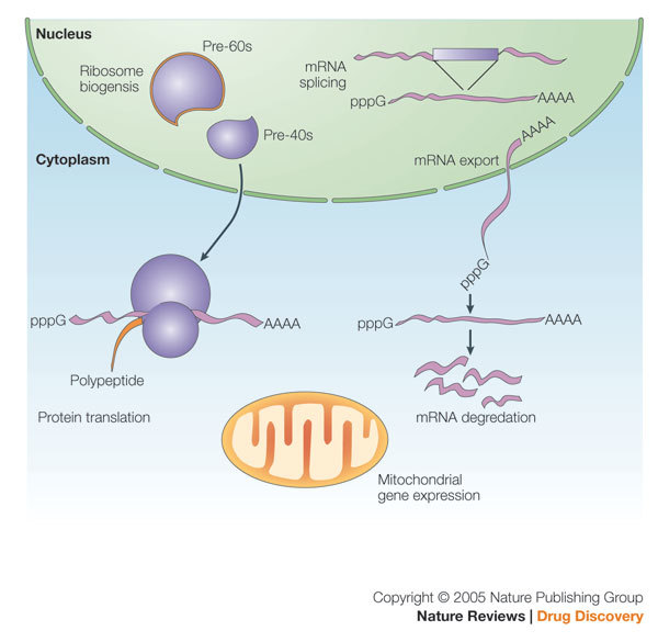
Various helicases have been implicated in transcription, mRNA splicing, mRNA transport, mRNA translation, ribosome biogenesis and mitochondrial gene expression.
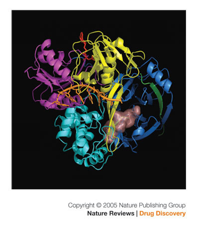
The three domains of the helicase in the front are coloured as in Fig. 1a. The amino-terminal protease domain is coloured blue. A protease inhibitor, VX-950 (coloured pink), is modelled into the active site of the protease at the interface of the carboxy-terminal third domain of the helicase. The poly(U) (orange) and ATP (red) were modelled into the structure by overlaying the full-length NS3 structure with the structures of helicase domain bound with poly(U) (PDB code: 1A1V ) and the PcrA helicase bound with ATP (PDB code: 1QHH ). It is clear from this picture that NS3 can function independently as a helicase and as a protease.
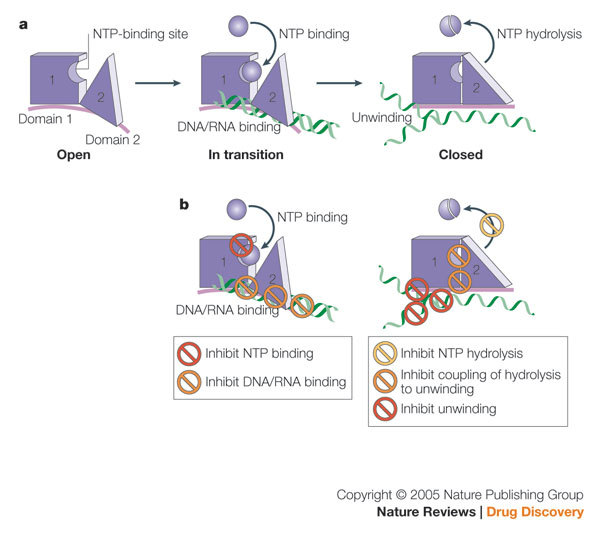
a | Schematic view of helicase reaction. b | Potential mechanisms of action for a small-molecule inhibitor. See discussion in text.
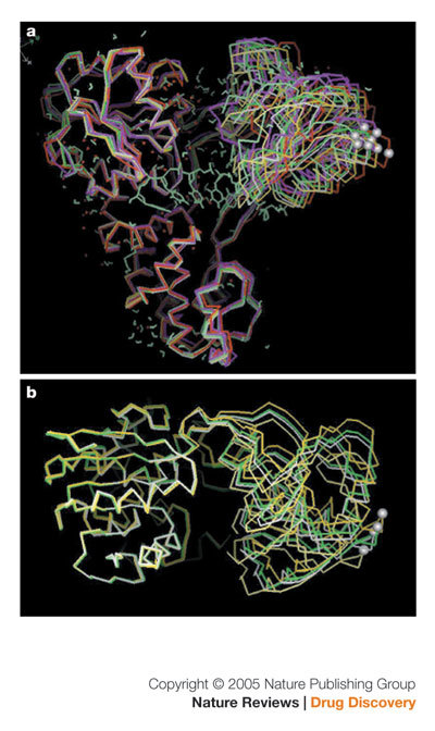
Superimposition of independent hepatitis C virus helicase structures. a | Superimposition of eight independently derived structures ('side view'). b | Superimposition of four independently derived structures ('top view').
References
-
- De Clercq E. Strategies in the design of antiviral drugs. Nature Rev. Drug Discov. 2002;1:13–25. - PubMed
-
- Richman DD. The implications of drug resistance for strategies of combination antiviral chemotherapy. Antiviral Res. 1996;29:31–33. - PubMed
-
- Lohman TM, Bjornson KP. Mechanisms of helicase-catalyzed DNA unwinding. Annu. Rev. Biochem. 1996;65:169–214. - PubMed
-
- Soultanas P, Wigley DB. Unwinding the 'Gordian knot' of helicase action. Trends Biochem. Sci. 2001;26:47–54. - PubMed
Publication types
MeSH terms
Substances
LinkOut - more resources
Full Text Sources
Other Literature Sources
