. 2005 Oct;115(10):2843-54.
doi: 10.1172/JCI25256. Epub 2005 Sep 22.

Polyunsaturated fatty acids suppress glycolytic and lipogenic genes through the inhibition of ChREBP nuclear protein translocation

Affiliations

Polyunsaturated fatty acids suppress glycolytic and lipogenic genes through the inhibition of ChREBP nuclear protein translocation

Renaud Dentin et al. J Clin Invest. 2005 Oct.

Abstract

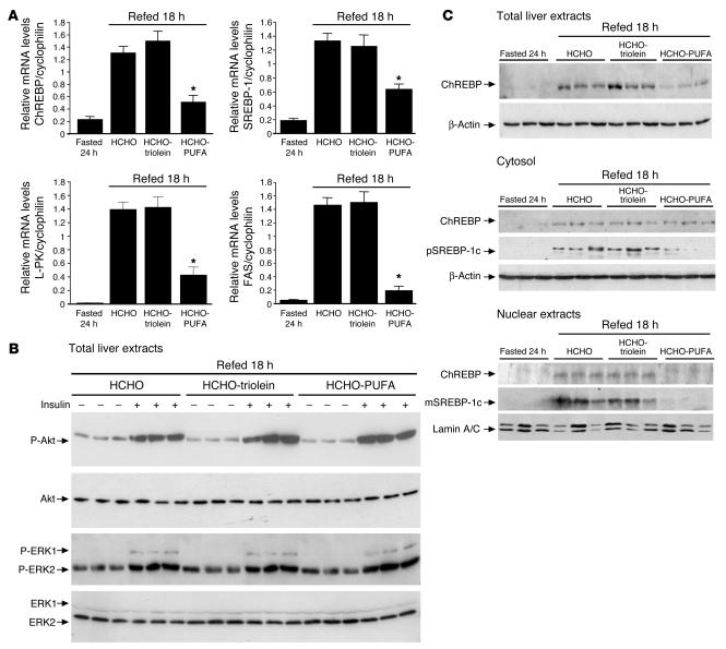
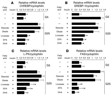
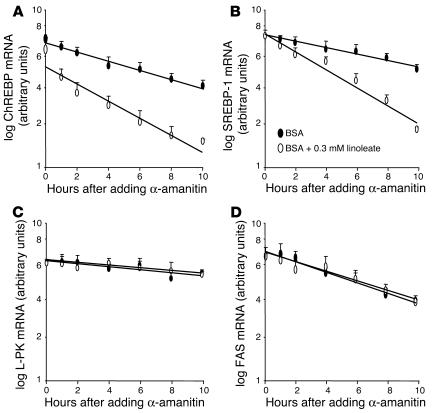
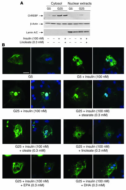
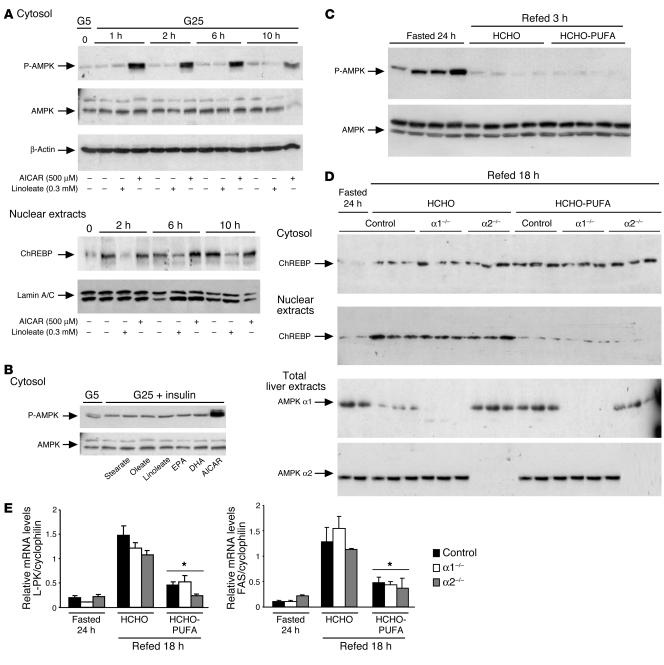
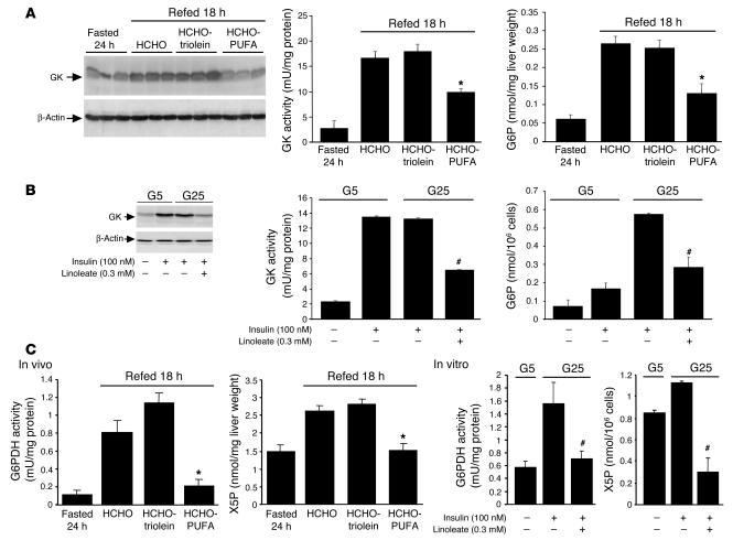
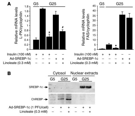
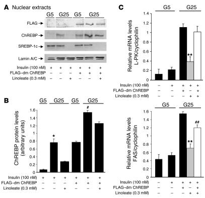
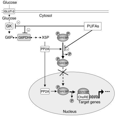
Dietary polyunsaturated fatty acids (PUFAs) are potent inhibitors of hepatic glycolysis and lipogenesis. Recently, carbohydrate-responsive element-binding protein (ChREBP) was implicated in the regulation by glucose of glycolytic and lipogenic genes, including those encoding L-pyruvate kinase (L-PK) and fatty acid synthase (FAS). The aim of our study was to assess the role of ChREBP in the control of L-PK and FAS gene expression by PUFAs. We demonstrated in mice, both in vivo and in vitro, that PUFAs [linoleate (C18:2), eicosapentanoic acid (C20:5), and docosahexaenoic acid (C22:6)] suppressed ChREBP activity by increasing ChREBP mRNA decay and by altering ChREBP translocation from the cytosol to the nucleus, independently of an activation of the AMP-activated protein kinase, previously shown to regulate ChREBP activity. In contrast, saturated [stearate (C18)] and monounsaturated fatty acids [oleate (C18:1)] had no effect. Since glucose metabolism via the pentose phosphate pathway is determinant for ChREBP nuclear translocation, the decrease in xylulose 5-phosphate concentrations caused by a PUFA diet favors a PUFA-mediated inhibition of ChREBP translocation. In addition, overexpression of a constitutive nuclear ChREBP isoform in cultured hepatocytes significantly reduced the PUFA inhibition of both L-PK and FAS gene expression. Our results demonstrate that the suppressive effect of PUFAs on these genes is primarily caused by an alteration of ChREBP nuclear translocation. In conclusion, we describe a novel mechanism to explain the inhibitory effect of PUFAs on the genes encoding L-PK and FAS and demonstrate that ChREBP is a pivotal transcription factor responsible for coordinating the PUFA suppression of glycolytic and lipogenic genes.


Figures

Figure 1
Figure 1
Dietary PUFAs suppress the hepatic abundance of ChREBP mRNA and ChREBP total and nuclear protein content. (A) RTQ-PCR analysis of glycolytic and lipogenic genes from livers of 24 hour–fasted mice and mice refed a HCHO-triolein or HCHO-PUFA diet for 18 hours were performed. Results are the mean ± SEM; n = 6/group. *Significantly different from mice refed a HCHO diet for 18 hours (P < 0.005). (B) Insulin-stimulated liver lysates from 18 hour HCHO-, HCHO-triolein–, and HCHO-PUFA–fed mice blotted with anti–phospho-Akt (P-Akt) and anti–phospho-MAPK antibodies. Blots were then stripped and reprobed for total Akt and MAPK. n = 3/group. (C) Total, cytosolic, and nuclear ChREBP, precursor SREBP-1 (pSREBP-1), and mature SREBP-1 (mSREBP-1) protein, in cytosolic and nuclear extracts from livers of 24 hour–fasted and 18 hour–refed mice on a HCHO diet supplemented or not with PUFAs. β-Actin and Lamin A/C antibodies were used as loading controls. A representative Western blot is shown. n = 6/group.
Figure 2
Figure 2
PUFAs inhibit ChREBP gene expression in cultured hepatocytes. After plating, hepatocytes were cultured for 24 hours in the presence of 5 mM glucose. Hepatocytes were then incubated for 24 hours in the presence of 5 or 25 mM glucose (G5 or G25) with 100 nM insulin and 100 nM dexamethasone in the presence or not of 0.3 mM of albumin-bound stearate (C18), oleate [C18:1(n-9)], linoleate [C18:2 (n-6)], EPA [C20:5 (n-3)], or DHA [C22:6 (n-3)]. ChREBP (A), SREBP-1 (B), L-PK (C), and FAS (D) gene expression were measured by RTQ-PCR. Results are mean ± SEM of values obtained from 3 to 8 independent cultures. *Significantly different from 25 mM glucose plus insulin (P < 0.05).
Figure 3
Figure 3
PUFAs accelerate ChREBP mRNA decay in cultured hepatocytes. After plating, hepatocytes were cultured for 48 hours in the presence of 25 mM glucose and 100 nM insulin. Hepatocytes were then treated with 0.3 mM albumin-bound linoleate (open symbols) or albumin alone (filled symbols) for 2 hours prior to the addition of the transcription inhibitor α-amanitin (15 μM final). The abundance of ChREBP (A), SREBP-1 (B), L-PK (C) and FAS (D) mRNA was determined by RTQ-PCR. Results are the mean ± SEM of values obtained from 3 independent cultures.
Figure 4
Figure 4
PUFAs suppress nuclear translocation of ChREBP in cultured hepatocytes. After plating, hepatocytes were cultured for 24 hours in the presence of 5 mM glucose. Hepatocytes were then incubated for 24 hours in the presence of 5 or 25 mM glucose with or without 100 nM insulin and 100 nM dexamethasone containing or not 0.3 mM of albumin-bound linoleate. (A) Cytosolic and nuclear forms of ChREBP were measured. Representative Western blots of 4 independent cultures are shown. (B) Representative images of subcellular localization of GFP-fused ChREBP under 5 mM glucose with or without 100 nM insulin; and 25 mM glucose plus 100 nM insulin with or without 0.3 mM of albumin-bound stearate (C18), oleate [C18:1 (n-9)], linoleate [C18:2 (n-6)], EPA [C20:5 (n-3)], or DHA [C22:6 (n-3)]. For each condition, hepatocyte nuclei were specifically stained using DAPI (right panels). Scale bar, 10 μM.
Figure 5
Figure 5
Effect of PUFAs and AICAR on AMPK activation and ChREBP localization. In vitro studies: After plating, hepatocytes were cultured for 24 hours in the presence of 5 mM glucose and 100 nM insulin. (A) Hepatocytes were then incubated with 25 mM glucose and 100 nM insulin in the presence or not of 0.3 mM of albumin-bound linoleate or 500 μM AICAR. After 1, 2, 6, and 10 hours, cytosolic form of phospho-AMPK and nuclear forms of ChREBP were measured. (B) After a 6-hour incubation period with 25 mM glucose, 100 nM insulin, and 0.3 mM of albumin-bound stearate (C18), oleate [C18:1 (n-9)], linoleate [C18:2 (n-6)], EPA [C20:5 (n-3)], DHA [C22:6 (n-3)], or 500 μM AICAR, the cytosolic form of P-AMPK protein was measured. Representative Western blots of 4 independent cultures are shown. In vivo studies: (C) The phosphorylation status of AMPK was measured in liver extracts from 24 hour–fasted mice refed for 3 hours on a HCHO or a HCHO-PUFA diet. n = 4/group. (D) Cytosolic and nuclear ChREBP content from livers of 24 hour–fasted control and AMPKα1–/– or AMPKα2–/– (α1–/– and α2–/–) mice refed 18 hours upon HCHO diet supplemented or not with PUFAs. A representative Western blot is shown; n = 3/group. (E) RTQ-PCR analysis of L-PK and FAS genes from livers of 24 hour–fasted mice and mice refed 18 hours on a HCHO or HCHO-PUFA diet were performed in control and AMPKα1–/– or AMPKα2–/– mice. Results are the mean ± SEM; n = 3/group. *Significantly different from mice refed a HCHO diet for 18 hours (P < 0.005).
Figure 6
Figure 6
Key steps of glycolysis and pentose phosphate pathway are decreased by PUFAs both in vivo and in vitro. In vivo studies: Mice were fasted for 24 hours and then refed 18 hours on a HCHO-triolein or HCHO-PUFA diet. In vitro studies: After plating, hepatocytes were cultured for 24 hours in the presence of 5 mM glucose. Hepatocytes were then incubated for 24 hours in the presence of 5 or 25 mM glucose with 100 nM insulin in the presence or not of 0.3 mM of albumin-bound linoleate [C18:2 (n-6)]. (A) GK protein content and activity and G6P concentrations in vivo. Results are presented as the mean ± SEM; n = 6/group. *Significantly different from mice refed a HCHO diet for 18 hours (P < 0.005). (B) GK protein content and activity and G6P concentrations were measured in vitro. Results are mean ± SEM of values obtained from 4 independent cultures. #Significantly different from 25 mM glucose plus insulin (P < 0.005). (C) G6PDH activity and X5P concentrations in vivo and in vitro. Results are presented as the mean ± SEM; n = 6/group. *Significantly different from mice refed a HCHO diet for 18 hours (P < 0.005). #Significantly different from 25 mM glucose plus insulin (P < 0.005).
Figure 7
Figure 7
Overexpression of SREBP-1c in cultured hepatocytes overrides the PUFA suppression of FAS but not of L-PK gene expression. After plating, hepatocytes were cultured for 24 hours in the presence of 5 mM glucose. Hepatocytes were then incubated with 5 or 25 mM glucose with the Ad-SREBP-1c at 1 PFU/cell in the presence or not of 0.3 mM of albumin-bound linoleate. (A) After 24 hours, L-PK and FAS gene expression was analyzed by RTQ-PCR. Results are the mean ± SEM of values obtained from 4 independent cultures. *Significantly different from conditions with insulin and no linoleate (P < 0.05). #Significantly different from conditions with Ad-SREBP-1c and no linoleate. (B) Cytosolic and nuclear forms of ChREBP and mature form of SREBP-1c were measured. A representative Western blot of 4 independent cultures is shown.
Figure 8
Figure 8
Overexpression of dm ChREBP in cultured hepatocytes counteracts the PUFA suppression of L-PK and FAS genes. After plating, hepatocytes were cultured for 24 hours in the presence of 5 mM glucose. Hepatocytes overexpressing dm ChREBP were then incubated with 5 or 25 mM glucose in the presence or not of 0.3 mM of albumin-bound linoleate. (A) Nuclear forms of ChREBP and the mature form of SREBP-1c were measured. A representative Western blot of 3 independent cultures is shown. (B) Quantification of ChREBP levels under the indicated experimental conditions. Results are the mean ± SEM of values obtained from 3 independent cultures. *Significantly different from 5 mM glucose plus insulin (P < 0.05). #Significantly different from FLAG–dm ChREBP at 5 mM glucose (P < 0.05). (C) After 24 hours, L-PK and FAS gene expression was measured by RTQ-PCR. Results are the mean ± SEM of values obtained from 3 independent cultures. **Significantly different from dm ChREBP in the presence of 25 mM glucose plus insulin (P < 0.05). ##Significantly different from 25 mM glucose plus insulin plus linoleate (P < 0.05).
Figure 9
Figure 9
Inhibitory effect of PUFAs on ChREBP activation and translocation. Under basal conditions of low glucose and insulin concentrations, ChREBP is phosphorylated and localized in the cytosol of hepatocytes. Its nuclear translocation is rapidly induced under high glucose and insulin concentrations. The nuclear translocation of ChREBP is controlled by a mechanism of dephosphorylation and phosphorylation. While the dephosphorylation of serine residue 196 (Ser196) allows ChREBP translocation into the nucleus, the dephosphorylation of threonine residue 666 (Thr666) alleviates DNA-binding inhibition. Protein phosphatase 2A (PP2A), selectively activated by X5P, is believed to be responsible for both cytosolic and nuclear dephosphorylation of ChREBP. Then ChREBP binds its response element (ChoRE) to activate glycolytic and lipogenic gene expression. In the presence of PUFAs, ChREBP is retained in the cytosol through the specific inhibition of GK and G6PDH activities, key enzymes of glycolysis and of the pentose phosphate pathway, respectively.

References

    1. Jump DB, Clarke SD. Regulation of gene expression by dietary fat. Annu. Rev. Nutr. 1999;19:63–90. - PubMed
    1. Liang G, et al. Diminished hepatic response to fasting/refeeding and liver X receptor agonists in mice with selective deficiency of sterol regulatory element-binding protein-1c. J. Biol. Chem. 2002;277:9520–9528. - PubMed
    1. Xu J, Teran-Garcia M, Park JH, Nakamura MT, Clarke SD. Polyunsaturated fatty acids suppress hepatic sterol regulatory element-binding protein-1 expression by accelerating transcript decay. J. Biol. Chem. 2001;276:9800–9807. - PubMed
    1. Worgall TS, Sturley SL, Seo T, Osborne TF, Deckelbaum RJ. Polyunsaturated fatty acids decrease expression of promoters with sterol regulatory elements by decreasing levels of mature sterol regulatory element-binding protein. J. Biol. Chem. 1998;273:25537–25540. - PubMed
    1. Hannah VC, Ou J, Luong A, Goldstein JL, Brown MS. Unsaturated fatty acids down-regulate srebp isoforms 1a and 1c by two mechanisms in HEK-293 cells. J. Biol. Chem. 2001;276:4365–4372. - PubMed

Publication types

MeSH terms