Comparative Study
. 2005 Oct 3;202(7):931-9.
doi: 10.1084/jem.20050715. Epub 2005 Sep 26.

Arginase I in myeloid suppressor cells is induced by COX-2 in lung carcinoma

Affiliations
Comparative Study

Arginase I in myeloid suppressor cells is induced by COX-2 in lung carcinoma

Paulo C Rodriguez et al. J Exp Med. .

Abstract

Myeloid suppressor cells (MSCs) producing high levels of arginase I block T cell function by depleting l-arginine in cancer, chronic infections, and trauma patients. In cancer, MSCs infiltrating tumors and in circulation are an important mechanism for tumor evasion and impair the therapeutic potential of cancer immunotherapies. However, the mechanisms that induce arginase I in MSCs in cancer are unknown. Using the 3LL mouse lung carcinoma, we aimed to characterize these mechanisms. Arginase I expression was independent of T cell-produced cytokines. Instead, tumor-derived soluble factors resistant to proteases induced and maintained arginase I expression in MSCs. 3LL tumor cells constitutively express cyclooxygenase (COX)-1 and COX-2 and produce high levels of PGE2. Genetic and pharmacological inhibition of COX-2, but not COX-1, blocked arginase I induction in vitro and in vivo. Signaling through the PGE2 receptor E-prostanoid 4 expressed in MSCs induced arginase I. Furthermore, blocking arginase I expression using COX-2 inhibitors elicited a lymphocyte-mediated antitumor response. These results demonstrate a new pathway of prostaglandin-induced immune dysfunction and provide a novel mechanism that can help explain the cancer prevention effects of COX-2 inhibitors. Furthermore, an addition of arginase I represents a clinical approach to enhance the therapeutic potential of cancer immunotherapies.


Figures

Figure 1.
Figure 1.
Expression of arginase I in tumor-infiltrating MSCs is independent of lymphocytes. (A) Single-cell suspensions from individual subcutaneous 3LL tumors excised from tumor-bearing C57BL/6 and C57BL/6 Prkdc scid mice (n = 15 per group) were tested for arginase I expression via Western blot analysis. Representative results of 10 tumors are shown. (B) Arginase I expression was tested in freshly isolated MSCs infiltrating individual 3LL tumors from C57BL/6 and C57BL/6 Prkdc scid tumor-bearing mice (n = 15 per group). Representative results from 6 tumors are shown.
Figure 2.
Figure 2.
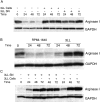
Arginase I expression in MSCs is induced by tumor-derived soluble factors. (A) MSCs (2 × 10⁶) isolated from 3LL tumors were cultured alone or cocultured in transwells with 2 × 10⁶ 3LL cells or 3LL supernatants. Arginase I expression was tested via Western blot analysis at 24, 48, and 72 h. (B) MSCs (2 × 10⁶) were cultured for 24 h in regular RPMI-1640, which results in the loss of arginase 1 expression. They were then cultured in RPMI-1640 alone or cocultured in transwells with 3LL cells (2 × 10⁶). Arginase I expression was tested at 24, 48, or 72 h afterward via Western blot analysis. (C) Peritoneal macrophages (2 × 10⁶) from normal mice were cocultured in transwells with 3LL cells (2 × 10⁶) or 3LL supernatants. Arginase I expression was tested via Western blot analysis. Results shown are representative of 3 experiments.
Figure 3.
Figure 3.
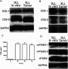
3LL cells and 3LL tumors express COX-1 and COX-2. 3LL cells cultured in vitro and single-cell suspension from subcutaneous tumors were tested for COX-1 and COX-2 protein (A) and mRNA (B). White lines indicate that intervening lanes have been spliced out. (C) Supernatants from 3LL cultured in vitro for 24 h were tested for PGE2 levels. (D) Cytoplasmic extracts from 3LL cells were also tested for PGES isoforms via Western blot analysis. Representative data are shown of experiments repeated three times.
Figure 4.
Figure 4.
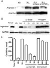
Arginase I induction in MSCs by 3LL tumor cells is dependent on COX-2. (A) MSCs (2 × 10⁶) were cultured in RPMI-1640 alone (NS or nonstimulated), 3LL supernatants, or 3LL supernatants containing 10 μg/ml anti-PGE2 or isotype control. Arginase I was tested 48 h later via Western blot analysis. (B, C) MSCs (2 × 10⁶) were cultured in RPMI 1640 (NS) or cocultured in transwells with 3LL cells (2 × 10⁶) in the presence of increasing concentrations (μM) of COX-1–specific (FR122047) and COX-2–specific (sc-58125) inhibitors. Arginase I expression was tested after 48 h of coculture via Western blot analysis, as was arginase activity via conversion of l-Arg to l-ornithine. *P < 0.0001 when comparing MSCs cocultured with 3LL with MSCs cocultured with 3LL in the presence of sc-58125 (20 μM).
Figure 5.
Figure 5.
COX-2 silencing in 3LL tumor cells blocks arginase I induction. (A) 3LL cells were transfected using COX-2 siRNA or an irrelevant siRNA probe as described in Material and methods. COX-2 mRNA expression was determined via Northern blot analysis. (B, C) After 24 h of transfection, 3LL cells (2 × 10⁶) were cocultured with freshly isolated MSCs (2 × 10⁶) using transwells, and arginase I expression and activity were tested in MSCs after 48 h. All experiments were repeated 3 times. *P < 0.005 when comparing MSCs cocultured with 3LL with MSCs cocultured with 3LL transfected with COX-2 siRNA.
Figure 6.
Figure 6.
EP4 activation induces arginase I in MSCs. (A) 3LL cells kept in culture and MSCs isolated from 3LL tumors were tested via Western blot analysis for EP receptor expression. (B, C) MSCs (2 × 10⁶) were cultured in the presence of 1 μM EP analogues for 24 h, after which arginase activity and arginase I expression were tested. White lines indicate that intervening lanes have been spliced out.
Figure 7.
Figure 7.
COX-2 inhibitor sc-58125 blocks arginase I induction in vivo. (A) Mice (n = 10) were injected subcutaneously with 3LL cells (1 × 10⁶) in the right flank and received subcutaneous injections of the COX-2 inhibitor sc-58125 (5 mg/kg) or DMSO (dilution vehicle) every other day for 18 d in the left flank. Tumor was excised and tumor single-cell suspensions were tested for arginase I and COX-2 via Western blot analysis. (B–D) Tumor volume was determined in tumor-bearing C57BL/6-Prkdc scid (n = 8), CD4−/− (n = 8), and CD8 −/− (n = 8) mice receiving sc-58125 or DMSO. (E) Single-cell suspensions from tumors were lysed and tested for VEGF and E-cadherin expression. One representative sample from eight different tumors is shown.
Figure 8.
Figure 8.
Arginase I is induced by tumor-derived COX-2. Arginase was tested in tumors harvested 18 d after tumor injection from COX-2 knockout mice. One representative sample of two different tumors is shown.

Similar articles

Cited by

References

    1. Gabrilovich, D.I., M.P. Velders, E.M. Sotomayor, and W.M. Kast. 2001. Mechanism of immune dysfunction in cancer mediated by immature Gr-1+ myeloid cells. J. Immunol. 166:5398–5406. - PubMed
    1. Bronte, V., P. Serafini, C. De Santo, I. Marigo, V. Tosello, A. Mazzoni, D.M. Segal, C. Staib, M. Lowel, G. Sutter, et al. 2003. IL-4-induced arginase 1 suppresses alloreactive T cells in tumor-bearing mice. J. Immunol. 170:270–278. - PubMed
    1. Liu, Y., J.A. Van Ginderachter, L. Brys, P. De Baetselier, G. Raes, and A.B. Geldhof. 2003. Nitric oxide-independent CTL suppression during tumor progression: association with arginase-producing (M2) myeloid cells. J. Immunol. 170:5064–5074. - PubMed
    1. Mazzoni, A., V. Bronte, A. Visintin, J.H. Spitzer, E. Apolloni, P. Serafini, P. Zanovello, and D.M. Segal. 2002. Myeloid suppressor lines inhibit T cell responses by an NO-dependent mechanism. J. Immunol. 168:689–695. - PubMed
    1. Rodriguez, P.C., D.G. Quiceno, J. Zabaleta, B. Ortiz, A.H. Zea, M.B. Piazuelo, A. Delgado, P. Correa, J. Brayer, E.M. Sotomayor, et al. 2004. Arginase I production in the tumor microenvironment by mature myeloid cells inhibits T-cell receptor expression and antigen-specific T-cell responses. Cancer Res. 64:5839–5849. - PubMed

Publication types

MeSH terms