. 2005 Sep 26;170(7):1029-37.
doi: 10.1083/jcb.200502129.

The Rac activator Tiam1 controls tight junction biogenesis in keratinocytes through binding to and activation of the Par polarity complex

Affiliations

The Rac activator Tiam1 controls tight junction biogenesis in keratinocytes through binding to and activation of the Par polarity complex

Alexander E E Mertens et al. J Cell Biol. .

Abstract

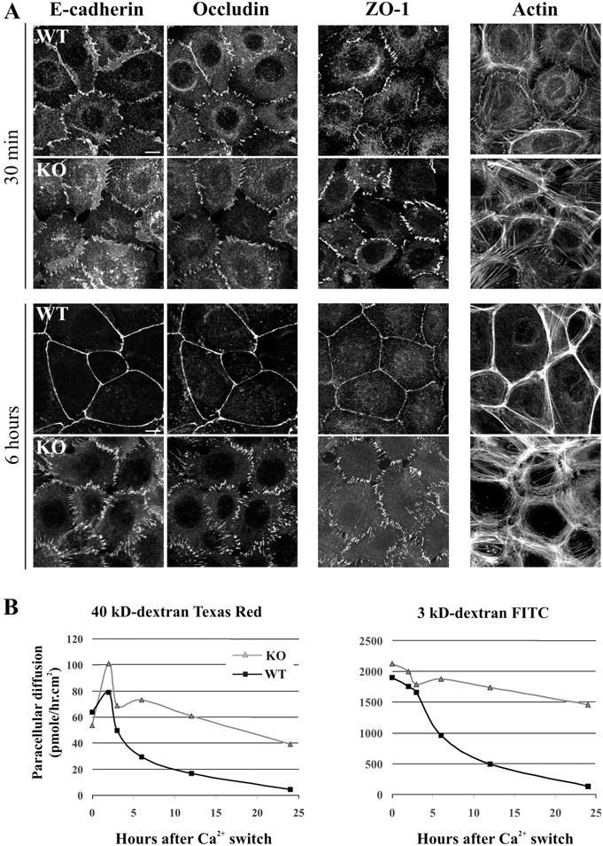
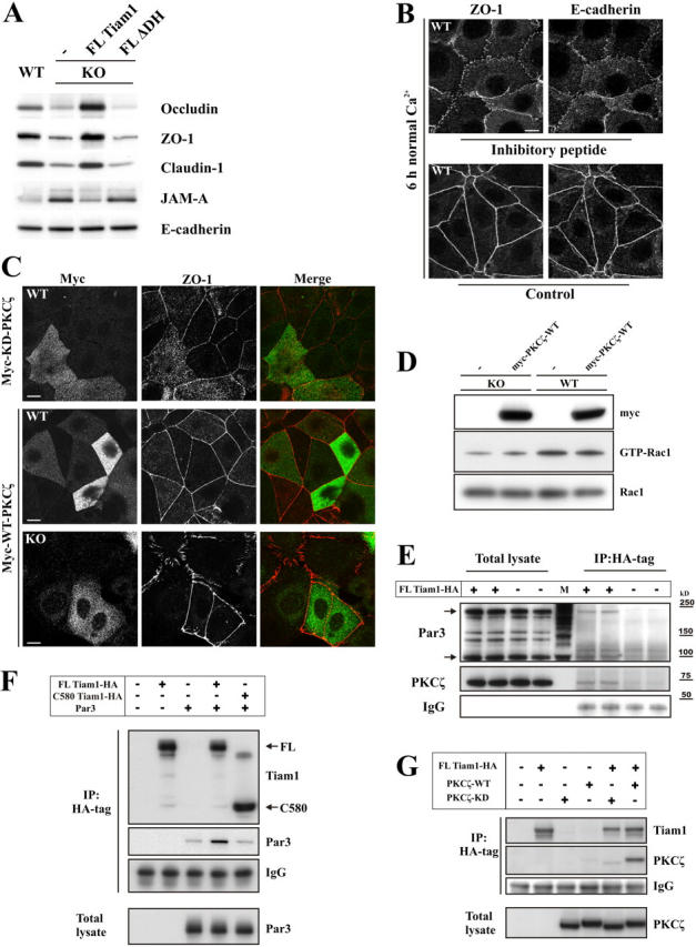
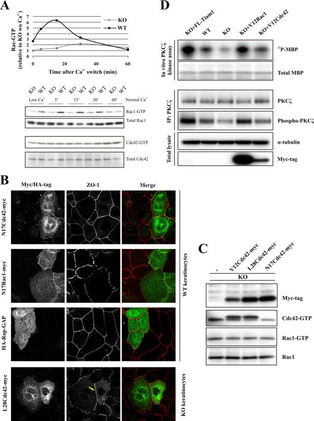
The GTPases Rac and Cdc42 play a pivotal role in the establishment of cell polarity by stimulating biogenesis of tight junctions (TJs). In this study, we show that the Rac-specific guanine nucleotide exchange factor Tiam1 (T-lymphoma invasion and metastasis) controls the cell polarity of epidermal keratinocytes. Similar to wild-type (WT) keratinocytes, Tiam1-deficient cells establish primordial E-cadherin-based adhesions, but subsequent junction maturation and membrane sealing are severely impaired. Tiam1 and V12Rac1 can rescue the TJ maturation defect in Tiam1-deficient cells, indicating that this defect is the result of impaired Tiam1-Rac signaling. Tiam1 interacts with Par3 and aPKCzeta, which are two components of the conserved Par3-Par6-aPKC polarity complex, and triggers biogenesis of the TJ through the activation of Rac and aPKCzeta, which is independent of Cdc42. Rac is activated upon the formation of primordial adhesions (PAs) in WT but not in Tiam1-deficient cells. Our data indicate that Tiam1-mediated activation of Rac in PAs controls TJ biogenesis and polarity in epithelial cells by association with and activation of the Par3-Par6-aPKC polarity complex.


Figures

Figure 1.
Figure 1.
Tiam1 is required for membrane sealing and TJ formation in epidermal keratinocytes. (A) Tiam1 was immunoprecipitated from cell lysates using anti-Tiam1 antibody (C16). Western blotting was performed with anti-Tiam1 antibody (DH). (B–D) WT and Tiam1KO cells were grown to confluency and incubated with 1.8 mM Ca2+ (normal Ca2+) for 6 h. (B) Phase-contrast images show sealing of the paracellular space in WT but not in Tiam1KO cells. Bars, 20 μm. (C) Paracellular diffusion of 3 kD FITC-dextran through keratinocyte monolayers that were cultured for 6 h in 1.8 mM Ca2+. Paracellular diffusion was measured for 2.5 h. Data are shown as means ± SD from three independent experiments. (D) EM images of the apical part of the lateral plasma membrane of keratinocyte monolayers. Arrowhead indicates TJ; arrows indicate desmosomes. Boxes with dotted lines indicate the area that is magnified in the bottom panel. Notice the membrane fusions that are typical of TJs in WT cells. Bars, 200 nm.
Figure 2.
Figure 2.
Tiam1 deficiency impairs junction maturation and TJ barrier function. (A) Tiam1 is not essential for the formation of PAs, but its absence impairs subsequent junction maturation. The concentration of Ca2+ was increased to 1.8 mM in the culture medium of keratinocyte monolayers for the indicated times. Cells were double stained with anti–E-cadherin and antioccludin antibodies or stained with anti–ZO-1 antibody or phalloidin for F-actin. Bars, 10 μm. (B) Tiam1 deficiency impairs TJ barrier function. Paracellular diffusion of 3 kD FITC-dextran and 40 kD Texas red–dextran through keratinocyte monolayers, which were cultured in 1.8 mM Ca2+ for the indicated time points. Paracellular diffusion was measured for 2.5 h. A representative example of three independent experiments is shown.
Figure 3.
Figure 3.
Tiam1 signaling to Rac is required for junction maturation and TJ formation. (A) Expression of V12Rac1 or FL-Tiam1, but not of catalytically inactive Tiam1 (Tiam1ΔDH), in Tiam1−/− cells restores junction maturation within 6 h after a Ca2+ switch. (B) Western blot analysis and Rac activity assays on total lysates from Tiam1KO cells stably expressing the indicated Tiam1 constructs and myc-tagged V12Rac1. (C) Expression of FL-Tiam1, but not Tiam1ΔDH, restores TJ barrier function in Tiam1KO keratinocytes. Paracellular diffusion was measured at the indicated time points after a Ca2+ switch. (D) Primary WT and Tiam1KO keratinocytes (4 d after isolation) that were switched to normal Ca2+ for 6 h display a similar phenotype as immortilized keratinocytes. Cells were double stained for ZO-1 and E-cadherin. (E) Down-regulation of Tiam1 disturbs junction maturation in WT keratinocytes. EGFP was cotransfected with pSUPER-siRNA-Tiam1 or with siRNA-luciferase (control) into WT keratinocytes in a molar ratio of 1:10. After 48 h, cells were stained for ZO-1 (6 h after the Ca2+ switch). Asterisks indicate GFP-positive (transfected) cells, as shown on the left. Bars, 10 μm.
Figure 4.
Figure 4.
Tiam1 regulates the expression of TJ molecules and interacts with the Par3–Par6–PKCζ polarity complex. (A) Tiam1–Rac signaling differentially regulates the expression of TJ molecules. Lysates of keratinocytes cultured in low Ca2+ medium were immunoblotted for the indicated junctional proteins. (B) Membrane-associated occludin facilitates junction maturation. WT cells were switched to 1.8 mM Ca2+ for 6 h in the presence or absence (DMSO control) of an occludin inhibitory peptide. (C) WT and Tiam1KO keratinocytes were transiently transfected with myc-tagged WT- or kinase-dead PKCζ-K281W, switched to normal Ca2+ for 6 h, fixed, and double stained for myc and ZO-1. PKCζ-K281W disturbs the junctional localization of ZO-1 in WT keratinocytes, whereas WT-PKCζ has no effect. WT-PKCζ fully restores junction maturation in Tiam1KO keratinocytes. Bars, 10 μm. (D) Rac1 activity assay on WT and Tiam1KO cells stably expressing myc–WT-PKCζ. (E–G) HA-tagged Tiam1 was expressed in Tiam1KO keratinocytes (E) or in COS-7 cells (F and G) by retroviral transduction and immunoprecipitated using anti-HA antibody. (E) FL-Tiam1 coimmunoprecipitates endogenous Par3 and aPKCζ from two independent infected populations of Tiam1KO cells that expressed HA–FL-Tiam1 and were cultured for 6 h at 1.8 mM Ca2+. Empty vector–infected Tiam1KO cells were used as a control. M, molecular mass marker. (F) FL-Tiam1, but not a COOH-terminal Tiam1 mutant (C580), containing the guanine nucleotide exchange factor domain only, coimmunoprecipitates Par3 from COS-7 cell lysates. (G) FL-Tiam1 coimmunoprecipitates exogenous WT- but not PKCζ-K281W from COS-7 cell lysates. Immunocomplexes were immunoblotted using the indicated antibodies.
Figure 5.
Figure 5.
Tiam1 and Rac1, but not Cdc42 or Rap1, control the activity of the polarity complex and TJ formation. (A) Cadherin signaling transiently activates Rac in a Tiam1-dependent manner. Rac and Cdc42 activity assays from WT and Tiam1KO keratinocytes after a Ca2+ switch for the indicated times. The graph represents the level of activated Rac (corrected for total Rac) relative to its activity in Tiam1KO cells cultured in low Ca2+. (B) WT keratinocytes were transiently transfected with myc-N17Cdc42, -N17Rac1, or HA-Rap1GAP. Tiam1KO keratinocytes were transfected with myc-L28Cdc42. 48 h after transfection, cells were switched to normal Ca2+ for 6 h, fixed, and double stained for myc or HA and ZO-1. Arrow indicates linearization of the TJ. (C) Tiam1, Rac, and Cdc42 can regulate PKCζ activity. Endogenous PKCζ was immunoprecipitated from WT, Tiam1KO, and Tiam1KO cells expressing FL-Tiam1, V12Rac1, or L28Cdc42. PKCζ kinase activity was assayed in vitro by using MBP as a substrate. Thr410 phosphorylation in the activation loop of PKCζ was analyzed as an alternative way to measure its activity. α-Tubulin was used as a loading control. Representative example of four experiments. (D) Cdc42 and Rac activity assay in Tiam1KO cells stably expressing myc-tagged L28, V12, or N17Cdc42 mutants.

References

    1. Arthur, W.T., L.A. Quilliam, and J.A. Cooper. 2004. Rap1 promotes cell spreading by localizing Rac guanine nucleotide exchange factors. J. Cell Biol. 167:111–122. - PMC - PubMed
    1. Chen, X., and I.G. Macara. 2005. Par-3 controls tight junction assembly through the Rac exchange factor Tiam1. Nat. Cell Biol. 7:262–269. - PubMed
    1. Fukuhara, A., K. Shimizu, T. Kawakatsu, T. Fukuhara, and Y. Takai. 2003. Involvement of nectin-activated Cdc42 small G protein in organization of adherens and tight junctions in Madin-Darby canine kidney cells. J. Biol. Chem. 278:51885–51893. - PubMed
    1. Gao, L., G. Joberty, and I.G. Macara. 2002. Assembly of epithelial tight junctions is negatively regulated by Par6. Curr. Biol. 12:221–225. - PubMed
    1. Itoh, M., H. Sasaki, M. Furuse, H. Ozaki, T. Kita, and S. Tsukita. 2001. Junctional adhesion molecule (JAM) binds to PAR-3: a possible mechanism for the recruitment of PAR-3 to tight junctions. J. Cell Biol. 154:491–497. - PMC - PubMed

Publication types