. 2005 Sep 26;170(7):1127-34.
doi: 10.1083/jcb.200503011.

Neurotransmitter release regulated by a MALS-liprin-alpha presynaptic complex

Affiliations

Neurotransmitter release regulated by a MALS-liprin-alpha presynaptic complex

Olav Olsen et al. J Cell Biol. .

Abstract

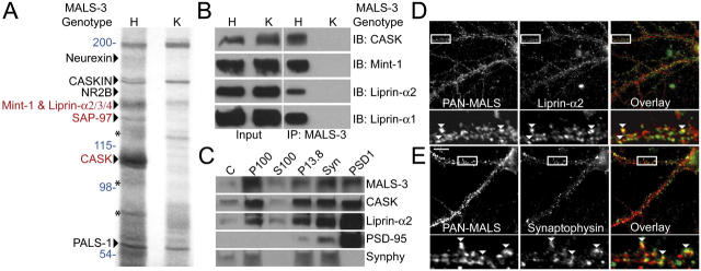
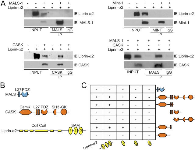
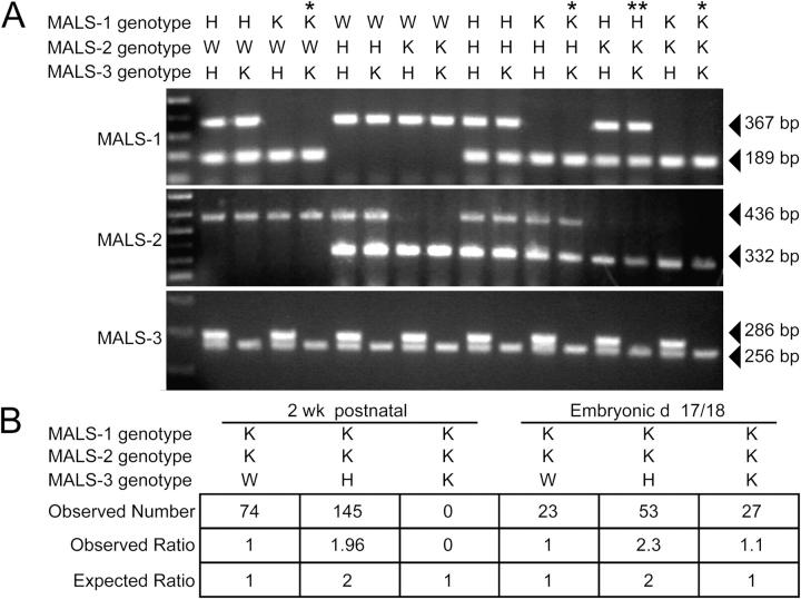
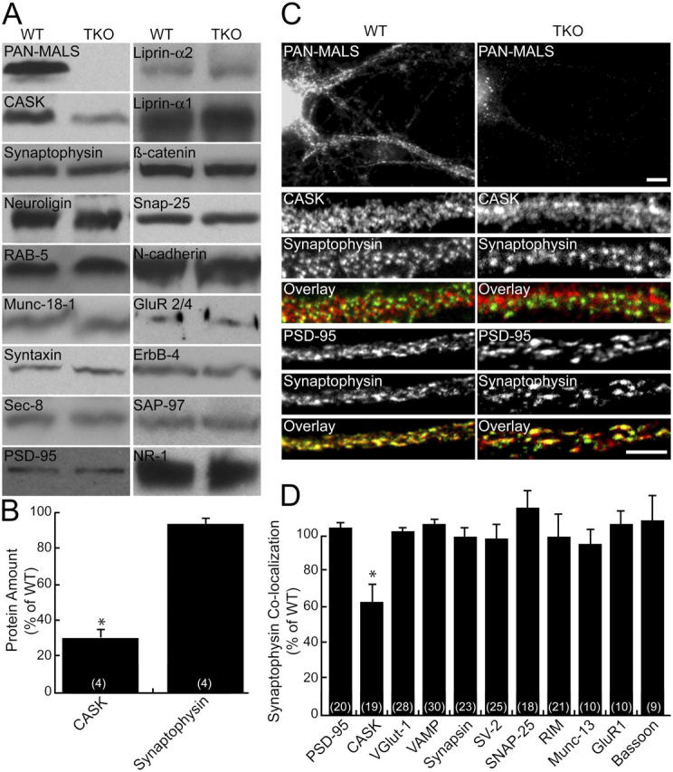
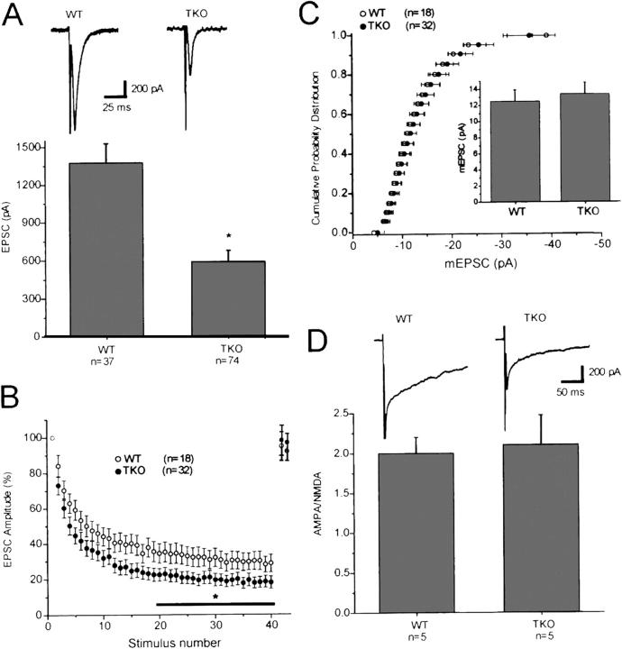
Synapses are highly specialized intercellular junctions organized by adhesive and scaffolding molecules that align presynaptic vesicular release with postsynaptic neurotransmitter receptors. The MALS/Veli-CASK-Mint-1 complex of PDZ proteins occurs on both sides of the synapse and has the potential to link transsynaptic adhesion molecules to the cytoskeleton. In this study, we purified the MALS protein complex from brain and found liprin-alpha as a major component. Liprin proteins organize the presynaptic active zone and regulate neurotransmitter release. Fittingly, mutant mice lacking all three MALS isoforms died perinatally with difficulty breathing and impaired excitatory synaptic transmission. Excitatory postsynaptic currents were dramatically reduced in autaptic cultures from MALS triple knockout mice due to a presynaptic deficit in vesicle cycling. These findings are consistent with a model whereby the MALS-CASK-liprin-alpha complex recruits components of the synaptic release machinery to adhesive proteins of the active zone.


Figures

Figure 1.
Figure 1.
Identification of a neuronal protein complex containing MALS and liprin-α. (A) Immunoprecipitation of MALS-3 from brain extracts showed a series of bands in heterozygote (H) that were absent from MALS-3 knockout (K). Bands were identified by MS/MS obtained using a micro-ionspray source attached to a mass spectrometer (red) and confirmed by Western blotting (black). Molecular weights are presented in blue and Mint-1 degradation products are shown with asterisks. (B) Western blotting of heterozygote and knockout brain extracts immunoprecipitated for MALS-3 shows specific association of CASK, Mint-1, liprin-α1, and -α2 with MALS-3. (C) Western blotting shows that MALS-3, CASK, and liprin-α2 are highly enriched with synaptophysin (Synphy) in the synaptosome (Syn) fraction and PSD-95 in PSD fractions. (D and E) Hippocampal cultures (28 DIV) were stained for MALS, liprin-α2, and synaptophysin. Immunostaining reveals that both liprin-α2 (D) and synaptophysin (E) partially colocalize with MALS (arrowheads). Bar, 20 μm.
Figure 2.
Figure 2.
Domain mapping of the MALS–liprin-α interaction. (A) CASK, but not MALS-1 or Mint-1, coimmunoprecipitated liprin-α2 in transfected COS cells. In the presence of CASK, MALS coimmunoprecipitated liprin-α2. (B) Schematics representing the structural domains of MALS-3 (blue), CASK (orange), and liprin-α2 (yellow). (C) By yeast two-hybrid analysis, CASK, but not MALS-3, interacted with liprin-α2. For CASK–liprin-α binding, the SAM1 domain of liprin-α2 is sufficient for interaction with CASK. Both the CaMK-like domain and first L27 domain of CASK were necessary for binding to liprin-α2.
Figure 3.
Figure 3.
Generation of mice lacking all three MALS isoforms. (A) PCR genotyping of MALS mice. Single asterisk indicates mice that died within hours of birth and double asterisk indicates a line that died in their second postnatal week. (B) Statistics obtained from crossing MALS-1/2 K and MALS-3 H mice. Genotyping 2-wk-old mice showed no TKO mice. However, embryonic mice (E18) showed the predicted ratio of W, H, and K mice.
Figure 4.
Figure 4.
CASK expression is reduced in MALS-deficient mice. (A) Brains from E18 mice were immunoblotted for numerous synaptic proteins. (B) CASK was markedly reduced (31% of control ± 8; *, P < 0.01) in the TKOs, but no changes in other synaptic proteins were detected. (C and D) Similarly, cultured hippocampal neurons lacking MALS displayed normal localization of several pre- and postsynaptic markers but showed reduced colocalization of CASK (red) and synaptophysin (green), suggesting that CASK is partially lost from synapses (62% ± 12 of control; *, P < 0.01). Bars: (top) 10 μm; (bottom) 5 μm. All error bars represent SEM.
Figure 5.
Figure 5.
A dominant-negative liprin-α disrupts presynaptic localization of MALS. Hippocampal cultures (35 DIV) were infected with Semliki Forest virus expressing either GFP or GFP fused to the SAM domains of liprin-α2. Whereas infection and expression of GFP had no effect on synaptic expression of MALS (A, arrowheads), expression of the dominant-negative (GFP-SAM) construct misdirected MALS to nonsynaptic sites (B, arrows) and resulted in a loss of presynaptic MALS (B, arrowheads). Quantification of immunofluorescence reveals that colocalization of MALS (red in overlayed images) with synaptophysin (green in overlayed images) is significantly reduced in neurons expressing GFP-SAM, from 77.3% ± 3.6 in uninfected neurons to 48.1% ± 2.1 and to 79.5% ± 2.0 in GFP-expressing neurons (P < 0.01). Bars: (top) 20 μm; (bottom) 10 μm.
Figure 6.
Figure 6.
Synapse size, but not number, is altered in neurons lacking MALS. Autaptic cultures (14 DIV) from control and TKO mice were stained with antibodies to PSD-95 and synaptophysin. (A) Representative autaptic neurons from WT and TKO mice stained with synaptophysin. Bars: (top) 20 μm; (bottom) 5 μm. (B) PSD-95/synaptophysin staining revealed that the number of synapses was unaltered in TKO cultures (0.65 ± 0.15 and 0.67 ± 0.18 synapses/μm for WT and TKO, respectively). (C) As determined by synaptophysin staining, the distribution of presynaptic terminal size was shifted to the right in the TKO (P < 0.01). All error bars represent SEM.
Figure 7.
Figure 7.
Neurons from MALS-deficient mice display abnormal synaptic transmission. (A) EPSCs recorded in autaptic cultures prepared from E18 MALS TKO mice were profoundly reduced relative to EPSCs recorded in WT cultures (*, P = 0.01). Typical EPSCs recorded in WT and MALS TKO pyramidal cell autapses are shown above. (B) Normalized synaptic responses during a 10-Hz (4 s) stimulus train. Decay of EPSCs during the high frequency train was greater in the MALS TKO autapses than in WT autapses (*, P < 0.05). EPSCs returned to baseline values within 5 s of the end of the stimulus train in both populations (n = 18 and 7 for WT and MALS TKO, respectively, for the recovery). (C) The distribution and amplitude of miniature EPSCs was the same in autapses prepared from WT and MALS TKO mice (P > 0.1). (D) The ratio of AMPA EPSCs to NMDA EPSCs was also similar in WT and MALS TKO autapses (P > 0.1). AMPA EPSCs were measured at the peak (∼8–10 ms after the action potential), whereas NMDA EPSCs were measured at 40 ms after the action potential, a time point when the AMPA EPSC has decayed to baseline. Typical AMPA/NMDA EPSCs from the two populations are shown. All error bars represent SEM.

References

    1. Augustin, I., C. Rosenmund, T.C. Südhof, and N. Brose. 1999. Munc13-1 is essential for fusion competence of glutamatergic synaptic vesicles. Nature. 400:457–461. - PubMed
    1. Bekkers, J.M., and C.F. Stevens. 1991. Excitatory and inhibitory autaptic currents in isolated hippocampal neurons maintained in cell culture. Proc. Natl. Acad. Sci. USA. 88:7834–7838. - PMC - PubMed
    1. Borg, J.-P., Y. Yang, M. De Taddéo-Borg, B. Margolis, and R.S. Turner. 1998. The X11alpha protein slows cellular amyloid precursor protein processing and reduces Abeta40 and Abeta42 secretion. J. Biol. Chem. 273:14761–14766. - PubMed
    1. Borg, J.-P., M.O. Lõpez-Figueroa, M. de Taddèo-Borg, D.E. Kroon, R.S. Turner, S.J. Watson, and B. Ben Margolis. 1999. Molecular analysis of the X11-mLin-2/CASK complex in brain. J. Neurosci. 19:1307–1316. - PMC - PubMed
    1. Butz, S., M. Okamoto, and T.C. Südhof. 1998. A tripartite protein complex with the potential to couple synaptic vesicle exocytosis to cell adhesion in brain. Cell. 94:773–782. - PubMed

Publication types

MeSH terms