. 2005 Oct;46(10):3623-30.
doi: 10.1167/iovs.05-0028.

Targeted deletion of AP-2alpha leads to disruption in corneal epithelial cell integrity and defects in the corneal stroma

Affiliations

Targeted deletion of AP-2alpha leads to disruption in corneal epithelial cell integrity and defects in the corneal stroma

Dhruva J Dwivedi et al. Invest Ophthalmol Vis Sci. 2005 Oct.

Abstract

Purpose: The present study was undertaken to create a conditional knockout of AP-2alpha in the corneal epithelium.

Methods: A line of mice expressing Cre-recombinase specifically in the early lens placode was crossed with mice in which the AP-2alpha allele is flanked by two loxP sites. The resultant Le-AP-2alpha mutants exhibited a targeted deletion of AP-2alpha in lens placode derivatives, including the differentiating corneal epithelium.

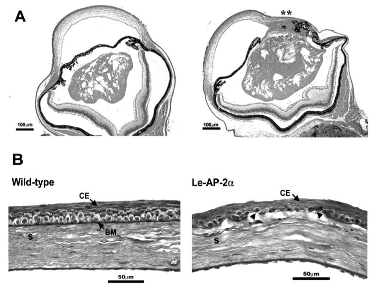
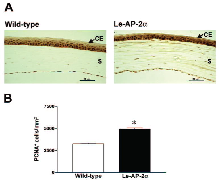
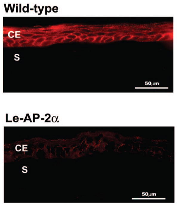
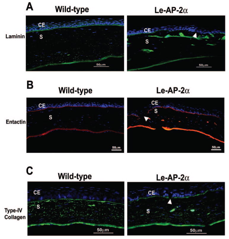
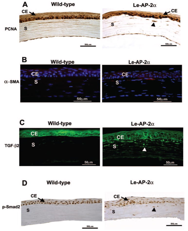
Results: The Le-AP-2alpha mutant mice were viable and had a normal lifespan. The adult corneal epithelium exhibited a variation in the number of stratified epithelial layers, ranging from 2 to 10 cell layers. A substantial decrease in expression of the cell-cell adhesion molecule, E-cadherin, was observed in all layers of the Le-AP-2alpha mutant corneal epithelium. The basement membrane, or Bowman's layer, was thinner in the mutant cornea and in many regions was discontinuous. These defects corresponded with altered distribution of laminin and entactin, and to a lesser degree, type IV collagen. The Le-AP-2alpha mutant cornea also exhibited stromal defects, including disrupted organization of the collagen lamellae and accumulation of fibroblasts beneath the epithelium that showed increased immunoreactivity for proliferating cell nuclear antigen (PCNA), alpha-smooth muscle actin (alpha-SMA), p-Smad2, and TGF-beta2.

Conclusions: In the absence of AP-2alpha, the corneal epithelium exhibits altered cell adhesion and integrity and defects in its underlying basement membrane. These defects likely caused the alterations in the corneal stroma.


Figures

Figure 1
Figure 1
Conditional knockout of AP-2α in the corneal epithelium. (A) PCR analysis of the Le-Cre-mediated deletion using the primers Alflp, Alflox4, and Alfscsq. The Le-AP-2α mouse tissues contain the AP-2α null allele and this generates a 490-bp product with primers Alflox4 and Alfscsq (E, ear; C, cornea). In addition, these tissues also contain the undeleted allele that generates a band of 560 bp with primers Alflox4 and Alfscsq. The deleted conditional allele produces a 185-bp band from primers Alflp and Alfscsq and is only detected in the cornea of Le-AP-2α mutants in the presence of Le-Cre. (B) Immunolocalization of AP-2α in the corneal epithelium of wild-type versus Le-AP-2α mutant mice. A FITC-conjugated secondary antibody was used to show specific staining for AP-2α. AP-2α was expressed in the more basally situated cells in the corneal epithelium of wild-type mice, whereas its expression was completely abolished in the corneal epithelium of the Le-AP-2α mutant mice. Some residual expression in the corneal epithelium is due to background in the Le-AP-2α mutant mice. CE, corneal epithelium; S, stroma.
Figure 2
Figure 2
(A) Hematoxylin and eosin-stained sections of adult Le-AP-2α mutant eyes. The mutant eye on the left exhibits a dysmorphic lens but no adhesion to the overlying cornea. The mutant eye on the right exhibits a severe adhesion of the lens to the peripheral cornea and the iris (**). (B) PAS staining of the basement membrane (Bowman's layer) in wild-type and Le-AP-2α mutant mouse corneas. The basement membranes of the corneas of wild-type mice were intact. In the Le-AP-2α mutant cornea, the basement membrane was thinner or absent (arrowheads) in some regions. CE, corneal epithelium, S, stroma.
Figure 3
Figure 3
(A) Immunostaining for PCNA in the corneal epithelium (CE) of wild-type and Le-AP-2α mutant mice. The corneal epithelium of Le-AP-2α mutant mice exhibited increased staining for PCNA in the suprabasal cell compartment compared with corneas of wild-type mice. (B) Statistical analysis of PCNA staining in the Le-AP-2α mutant versus wild-type corneal epithelium. The Le-AP-2α corneal epithelium showed a significant increase in the number of PCNA-positive corneal epithelial cells compared with wild-type littermates (n = 6; *P < 0.05). S, stroma.
Figure 4
Figure 4
Immunolocalization of E-cadherin in wild-type and Le-AP-2α mutant mouse corneas. E-cadherin staining (red, rhodamine-conjugated antibody) was observed along the cell–cell borders in all the stratified layers of the corneal epithelium (CE) of wild-type mice. In the Le-AP-2α mutant corneal epithelium, E-cadherin expression was substantially decreased. The integrity of the corneal epithelial layers was also altered in the Le-AP-2α mutant corneas. S, stroma.
Figure 5
Figure 5
Immunolocalization of basement membrane components (A) laminin, (B) entactin, and (C) type IV collagen in the Le-AP-2α mutant versus wild-type cornea. (A) Laminin was detected in the basement membrane underlying the corneal epithelium (CE) and the endothelium of wild-type and Le-AP-2α mutant mice by immunofluorescence with a FITC-conjugated secondary antibody (green). Note the discontinuous laminin staining (arrowhead) in the Le-AP-2α-mutant corneas and appearance of large aggregates of laminin in the anterior region of the stroma (S) in the Le-AP-2α mutant corneas. (B) Immunolocalization of entactin was observed in the basement membrane underlying the corneal epithelium and the endothelium of wild-type and Le-AP-2α mutant corneas using a rhodamine-conjugated secondary antibody (red). Note the discontinuous entactin staining in the Le-AP-2α mutant corneas with aggregates of entactin in the stroma (arrowhead). (C) Type IV collagen was localized with an FITC-conjugated secondary antibody (green). Type IV collagen was localized as a band of intense staining in the basement membrane underlying the corneal epithelium and the endothelium of both the wild-type and Le-AP-2α mutant corneas; however, note the small breaks in staining in the mutant (arrowhead). In all panels, the nuclei were colocalized by staining with DAPI (blue).
Figure 6
Figure 6
Markers of fibroblast activation are evident in the Le-AP-2α corneal stroma (S). (A) The proliferating status of the corneal stromal cells was determined by immunostaining for PCNA. Little to no proliferative response was observed in the stroma of the wild-type cornea. In contrast, stromal fibroblasts (arrowhead) in the Le-AP-2α mutant cornea exhibited immunoreactivity for PCNA, particularly in regions where the basement membrane was absent. (B) The activated response was further examined by immunofluorescent localization of α-SMA, with a CY3-conjugated secondary antibody (red). Cell nuclei were stained with DAPI (blue) to delineate the location of cells. The wild-type corneas did not exhibit any α-SMA protein expression, whereas some fibroblasts beneath the epithelium in the Le-AP-2α mutant corneas showed immunoreactivity to α-SMA. (C) TGF-β2 was immunolocalized with a FITC-conjugated secondary antibody (green). In wild-type mice TGF-β2 staining was absent in the corneal stroma and only evident in the corneal epithelium (CE). In the Le-AP-2α mutant cornea, both the corneal epithelium and stroma (S; arrowhead) stained positively for TGF-β2 (green). (D) Immunostaining for phospho-Smad2 (p-Smad2), a downstream effector of TGF-β2 signaling, showed that in wild-type mice, p-Smad2 was localized only in the epithelium, whereas in the Le-AP-2α mutant cornea both the stroma (arrowhead) and epithelium showed immunoreactivity to p-Smad2.

References

    1. Arffa RC. Grayson's Diseases of the Cornea. St. Louis, MO: Mosby; 1998.
    1. Trainor PA, Tam PP. Cranial paraxial mesoderm and neural crest cells of the mouse embryo: codistribution in the craniofacial mesenchyme but distinct segregation in branchial arches. Development. 1995;121:2569–2582. - PubMed
    1. Fini ME, Strissel KJ, West-Mays JA. Perspectives on eye development. Dev Genet. 1997;20:175–185. - PubMed
    1. Chow RL, Lang RA. Early eye development in vertebrates. Annu Rev Cell Dev Biol. 2001;17:255–296. - PubMed
    1. Hay ED. Development of the vertebrate cornea. Int Rev Cytol. 1979;63:263–322. - PubMed

Publication types

MeSH terms