. 2005 Nov 8;102(45):16133-40.
doi: 10.1073/pnas.0507057102. Epub 2005 Sep 26.

A bacterial group II intron-encoded reverse transcriptase localizes to cellular poles

Affiliations

A bacterial group II intron-encoded reverse transcriptase localizes to cellular poles

Junhua Zhao et al. Proc Natl Acad Sci U S A. .

Abstract

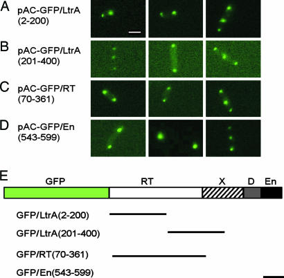
The Lactococcus lactis Ll.LtrB group II intron encodes a reverse transcriptase (LtrA protein) that binds the intron RNA to promote RNA splicing and intron mobility. Here, we used LtrA-GFP fusions and immunofluorescence microscopy to show that LtrA localizes to cellular poles in Escherichia coli and Lactococcus lactis. This polar localization occurs with or without coexpression of Ll.LtrB intron RNA, is observed over a wide range of cellular growth rates and expression levels, and is independent of replication origin function. The same localization pattern was found for three nonoverlapping LtrA subsegments, possibly reflecting dependence on common redundant signals and/or protein physical properties. When coexpressed in E. coli, LtrA interferes with the polar localization of the Shigella IcsA protein, which mediates polarized actin tail assembly, suggesting competition for a common localization determinant. The polar localization of LtrA could account for the preferential insertion of the Ll.LtrB intron in the origin and terminus regions of the E. coli chromosome, may facilitate access to exposed DNA in these regions, and could potentially link group II intron mobility to the host DNA replication and/or cell division machinery.


Figures

Fig. 1.
Fig. 1.
LtrA–GFP fusions are active in intron mobility. (A) Intron mobility assay. The CamR intron-donor plasmid pACD2X, which expresses a 0.9-kb Ll.LtrB-ΔORF intron with a phage T7 promoter near its 3′ end plus LtrA protein downstream of the 3′ exon, is cotransformed into E. coli HMS174(DE3) with the AmpR recipient plasmid pBRR3-ltrB. The latter contains the Ll.LtrB target site (positions –30 to +15) cloned upstream of a promoterless tetR gene. After induction with IPTG, insertion of the intron carrying the T7 promoter into the target site activates the tetR gene, and mobility frequencies are measured from the ratio of (TetR + AmpR)/AmpR colonies. PT7lac is the T7lac promoter used for donor intron expression, and T1, T2, and TΦ are transcription terminators for E. coli RNA polymerase (T1 and T2) and phage T7 RNA polymerase (TΦ). (B) Mobility assays with donor plasmids in which GFP is fused in-frame to LtrA's N terminus (GFP/LtrA) or C terminus (LtrA/GFP). The bar graphs show the mean ± SD for three experiments.
Fig. 2.
Fig. 2.
LtrA–GFP fusions are pole-localized in E. coli. Images show fluorescence microscopy. E. coli HMS174(DE3) containing the indicated plasmids was grown and induced with 250 μM IPTG for 1 h at 30°C. In E, DAPI and FM4–64 were added to stain DNA and cell membranes, respectively. Constructs are diagrammed to the right. The Ll.LtrB-ΔORF intron is indicated by an open rectangle with flanking exons shaded black. Deletions relative to the pACD2X–GFP/LtrA parent construct are indicated by breaks. MBP, maltose-binding protein. (Scale bar ≈ 2 μm; in E, magnification is ×1.3 that in other images.)
Fig. 3.
Fig. 3.
Immunofluorescence microscopy. E. coli HMS174(DE3) containing pACD2X was induced with 250 μM IPTG for 2 h at 37°C. After fixation, LtrA protein was detected with anti-LtrA antibody, followed by goat anti-rabbit IgG-FITC secondary antibody. (Scale bar ≈ 2 μm.)
Fig. 4.
Fig. 4.
LtrA is pole-localized in L. lactis.(A and B) Fluorescence microscopy. L. lactis NZ9800 containing pLE-RIG–GFP/LtrA or pLE-RIG–GFP, respectively, was grown and induced with nisin. (A) GFP fluorescence is superimposed over phase contrast images. (C) Detection of LtrA expressed from the endogenous Ll.LtrB intron in L. lactis NZ9800 by immunofluorescence microscopy. (D) pLE-RIG–GFP/LtrA contains the Ll.LtrB intron and short flanking exons (E1 and E2) cloned downstream of an inducible nisA promoter (PnisA). The ORF encoding the GFP/LtrA fusion is located in intron domain IV just upstream of a kanR–RIG marker (8). (Scale bar ≈ 2 μm.)
Fig. 5.
Fig. 5.
Localization of GFP fusions with different subsegments of LtrA. (AD) Fluorescence microscopy. E. coli HMS174(DE3) containing derivatives of pAC–GFP/LtrA with GFP fused to different subsegments of LtrA was induced with 250 μM IPTG at 37°C for 1 h. Synthesis of the correct-sized protein was confirmed by SDS/PAGE and Coomassie blue staining (AC) or by SDS/PAGE and immunoblotting with anti-GFP antibody (D). (E) Schematic of the LtrA protein with segments used in the GFP fusions delineated below. (Scale bar ≈ 2 μm.)
Fig. 6.
Fig. 6.
Polar localization of LtrA is not dependent on oriC function. (AD) Fluorescence microscopy. E. coli HMS174(DE3) containing pACD2X–GFP/LtrA (oriC+) (A) or AQ10033(DE3) oriC+ (B) and AQ10060(DE3) oriC (C and D) containing pACSD2–GFP/LtrA were induced with 250 μM IPTG for 1 h at 30°C. (C and D) Examples of AQ10060(DE3) oriC cells with normal and filamentous morphology. (E) Diagram of the oriC region in E. coli AQ10033 and AQ10060 in which the minimal oriC is replaced by an ampR gene. (Scale bar ≈ 2 μm.)
Fig. 7.
Fig. 7.
LtrA and IcsA may use related localization mechanisms. (AD) Competition experiments with E. coli HMS174(DE3) expressing IcsA507–620–GFP (pBAD24-icsA507–620::gfp) (A), IcsA507–620–GFP (pBAD24-icsA507–620::gfp) plus LtrA (pACD2X) (B), GFP/LtrA (placGFP/LtrA) (C), and GFP/LtrA (placGFP/LtrA) plus IcsA with the signal peptide deleted (pACD2X-IcsAΔSP) (D). Cells were induced with 0.2% l-arabinose (pBAD promoter) and/or 250 μM IPTG (T7lac promoter) for 4 h at 37°C. (E) Effect of aztreonam. E. coli HMS174(DE3) containing pAC–GFP/LtrA was grown in LB medium containing chloramphenicol and induced with 250 μM IPTG in the presence of 1 μg/ml aztreonam for 2 h at 37°C. DAPI was added to stain DNA. (F) Effect of DNase I on GFP/LtrA localization. HMS174(DE3) containing pACD2X–GFP/LtrA was induced with 250 μM IPTG at 37°C for 2 h and stained with DAPI. A portion of the cells was incubated with 100 μg/ml lysozyme (Sigma) and 100 units/ml DNase I (Invitrogen) for 1 h at room temperature before fluorescence microscopy. (Scale bar ≈ 2 μm.)

Similar articles

Cited by

References

    1. Belfort, M., Derbyshire, V., Parker, M. M., Cousineau, B. & Lambowitz, A. M. (2002) in Mobile DNA II, eds. Craig, N. L., Craigie, R., Gellert, M. & Lambowitz, A. M. (Am. Soc. Microbiol., Washington, DC), pp. 761–783.
    1. Lambowitz, A. M. & Zimmerly, S. (2004) Annu. Rev. Genet. 38, 1–35. - PubMed
    1. Cui, X., Matsuura, M., Wang, Q., Ma, H. & Lambowitz, A. M. (2004) J. Mol. Biol. 340, 211–231. - PubMed
    1. Blocker, F. J., Mohr, G., Conlan, L. H., Qi, L., Belfort, M. & Lambowitz, A. M. (2005) RNA 11, 14–28. - PMC - PubMed
    1. San Filippo, J. & Lambowitz, A. M. (2002) J. Mol. Biol. 324, 933–951. - PubMed

Publication types

MeSH terms

Substances

LinkOut - more resources