Comparative Study
. 2005 Oct 11;102(41):14771-6.
doi: 10.1073/pnas.0506489102. Epub 2005 Sep 26.

A positive feedback loop of phosphodiesterase 3 (PDE3) and inducible cAMP early repressor (ICER) leads to cardiomyocyte apoptosis

Affiliations
Comparative Study

A positive feedback loop of phosphodiesterase 3 (PDE3) and inducible cAMP early repressor (ICER) leads to cardiomyocyte apoptosis

Bo Ding et al. Proc Natl Acad Sci U S A. .

Abstract

cAMP plays crucial roles in cardiac remodeling and the progression of heart failure. Recently, we found that expression of cAMP hydrolyzing phosphodiesterase 3A (PDE3A) was significantly reduced in human failing hearts, accompanied by up-regulation of inducible cAMP early repressor (ICER) expression. Angiotensin II (Ang II) and the beta-adrenergic receptor agonist isoproterenol (ISO) also induced persistent PDE3A down-regulation and concomitant ICER up-regulation in vitro, which is important in Ang II- and ISO-induced cardiomyocyte apoptosis. We hypothesized that interactions between PDE3A and ICER may constitute an autoregulatory positive feedback loop (PDE3A-ICER feedback loop), and this loop would cause persistent PDE3A down-regulation and ICER up-regulation. Here, we demonstrate that ICER induction repressed PDE3A gene transcription. PDE3A down-regulation activated cAMP/PKA signaling, leading to ICER up-regulation via PKA-dependent stabilization of ICER. With respect to Ang II, the initiation of the PDE3A-ICER feedback loop depends on activation of Ang II type 1 receptor (AT1R), classical PKC(s), and CREB (cAMP response element binding protein). We further show that the PDE3A-ICER feedback loop is essential for Ang II-induced cardiomyocyte apoptosis. ISO and PDE3 inhibitors also induced the PDE3A-ICER feedback loop and subsequent cardiomyocyte apoptosis, highlighting the importance of this PDE3A-ICER feedback loop and cAMP signaling in cardiomyocyte apoptosis. Our findings may provide a therapeutic paradigm to prevent cardiomyocyte apoptosis and the progression of heart failure by inhibiting the PDE3A-ICER feedback loop.


Figures

Fig. 1.
Fig. 1.
Effects of Ang II on PDE3A and ICER expression in vivo and in vitro. (A) Western blots showing PDE3A and iCER protein expression in rat hearts with continous Ang II infusion for 7 days. (B) Cardiomyocyte apoptosis in Ang II-infused rat hearts measured by in situ TUNEL staining. (C) Western blots showing the effects of Ang II washout on PDE3A and ICER expression. Rat neonatal cardiomyocytes were exposed to Ang II (200 nM) for 12 h, followed by Ang II washout for indicated time. (D) Western blot showing the effects of AT1R inhibition before or after Ang II treatment on PDE3A and ICER expression. Cardiomyocytes were exposed to Ang II for indicated time with or without the AT1R selective antagonist losartan (10 μM). Data presented are from a representative experiment of at least two independent experiments.
Fig. 2.
Fig. 2.
Role of ICER on PDE3A gene down-regulation. (A) Western blots showing the effect of ICER overexpression on PDE3A protein levels. Rat neonatal cardiomyocytes were transduced with indicated amount of adenovirus carrying wild-type ICER (Ad-ICER) for 48 h. MOI represents multiplicity of infection. (B) Gel image showing the effect of ICER overexpression on PDE3A mRNA levels. Adenovirus transduced cardiomyocytes were analyzed for PDE3A mRNA levels by relative quantitative RT-PCR. (C) VISTA Plot of the 5′-flanking region of PDE3A gene detailing conserved regions between human and mouse, peaks represent conserved regions. The position of the first exon is gray. (D) Luciferase activity of PDE3A promoter constructs. Data presented are from a representative experiment of at least three independent experiments.
Fig. 3.
Fig. 3.
Effects of antisense ICER on Ang II-mediated PDE3A and ICER expression. Cardiomyocytes were transduced with Ad-LacZ (30 MOI) or Ad-ICER-AS (30 MOI) before (A) or after (B) Ang II stimulation at 200 nM for indicated time. PDE3A, ICER, and β-actin protein levels were measured by Western blotting. Data presented are from a representative experiment of at least two independent experiments.
Fig. 4.
Fig. 4.
Role of PDE3A enzymatic activity in the regulation of ICER and PDE3A expression. (A) Western blots showing the effect of PDE3A antisense (PDE3AAS) on ICER expression. Cardiomyocytes were transduced with Ad-PDE3A-AS for 48 h with the indicated amounts of virus. (B) Western blots showing time-dependent effects of milrinone (10 μM). (C) Gel image showing the effect of milrinone on PDE3A mRNA levels analyzed by relative quantitative RT-PCR. (D) Western blots showing the roles of wild-type PDE3A1 and catalytically inactive PDE3A1 [PDE3A1(mut)] in Ang II-induced changes of PDE3A and ICER expression. Cardiomyocytes were transduced with Ad-LacZ, AdPDE3A1, or Ad-PDE3A(mut) at 30 MOI for 24 h, followed by Ang II (200 nM) treatment for 48 h. (E) Western blots showing the roles of PKA, CREB, and ICER on milrinone-induced PDE3A and ICER expression. Cardiomyocytes were transduced with 30 MOI Ad-LacZ, Ad-PKI, Ad-CREB-DN, or Ad-ICER-AS for 24 h, followed with or without milrinone (10 μM) for 48 h. Data presented are from a representative experiment of at least two independent experiments.
Fig. 5.
Fig. 5.
Role of classical PKC, PKA, and CREB in initiating and maintaining the persistent PDE3A down-regulation and ICER induction. Western blots showing the effects of classic PKC inhibitor, PKI, and DN-CREB on PDE3A and ICER expression in cardiomyocytes are shown. Rat neonatal cardiomyocytes were treated with myristoylated PKCβ C2-4 inhibitor (10 μM) or transduced with Ad-LacZ, Ad-PKI, or Ad-DN-CREB at 30 MOI before (A and C) or after (B and D) Ang II (200 nM) stimulation for indicated time. Data presented are from a representative experiment of at least three independent experiments.
Fig. 6.
Fig. 6.
Role of PKA activation in ICER protein stability. (A) Schematic diagram showing experimental protocol. Cardiomyocytes were stimulated with Ang II for 24 h, transduced with Ad-LacZ, Ad-DN-CREB, or Ad-PKI for 6 h, and then treated with the protein synthesis inhibitor cycloheximide (10 μg/ml) for 2, 6, or 12 h. (B) Western blots showing ICER protein expression in cardiomyocytes treated with the above protocol. Data presented are from a representative experiment of at least two independent experiments.
Fig. 7.
Fig. 7.
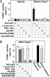
Role of the PDE3A-ICER feedback loop on Ang II-induced cardiomyocyte apoptosis. TUNEL staining data showing the effects of inhibiting AT1R, classical PKC, PKA, CREB, or ICER before (A) or after (B) Ang II stimulation, as well as Ang II washout on cardiomyocyte apoptosis (B), are shown. Cardiomyocytes were treated with AT1R antagonist losartan (10 μM) or myristoylated PKCβ C2-4 inhibitor (10 μM), or transduced with Ad-LacZ, Ad-PKI, Ad-DN-CREB, or Ad-ICER-AS at 30 MOI before (A) or 24 h after (B) Ang II (200 nM) treatment. Apoptosis was measured 48 h after Ang II treatment by TUNEL staining. For Ang II washout, cardiomyocytes were exposed to Ang II for 12 h, followed by Ang II washout for 48 h. Data represent mean of three repeats (mean ± SD). Similar results were obtained from at least two independent experiments.
Fig. 8.
Fig. 8.
Scheme of the PDE3A-ICER feedback loop Ang II, ISO, and PDE3 inhibitors induced sustained down-regulation of PDE3A expression and up-regulation of ICER, which is regulated by PDE3A-ICER positive feedback loop. In respect to Ang II, activation of PKC and CREB by Ang II via AT1R initiates the PDE3A-ICER feedback loop probably by inducing ICER gene transcription (dashed lines). Induction of ICER leads to a reduction of PDE3A expression and reduction of PDE3A leads to ICER elevation due to PKA-dependent stabilizing ICER protein from degradation, which constitutes the positive PDE3A-ICER feedback loop (solid lines) and allows a persistent induction of ICER that plays a key role in cardiomyocyte apoptosis.

Comment in

Similar articles

Cited by

References

    1. Kang, P. M. & Izumo, S. (2000) Circ. Res. 86, 1107-1113. - PubMed
    1. Olivetti, G., Abbi, R., Quaini, F., Kajstura, J., Cheng, W., Nitahara, J. A., Quaini, E., Di Loreto, C., Beltrami, C. A., Krajewski, S., Reed, J. C. & Anversa, P. (1997) N. Engl. J. Med. 336, 1131-1141. - PubMed
    1. Lohse, M. J., Engelhardt, S. & Eschenhagen, T. (2003) Circ. Res. 93, 896-906. - PubMed
    1. Leri, A., Liu, Y., Li, B., Fiordaliso, F., Malhotra, A., Latini, R., Kajstura, J. & Anversa, P. (2000) Am. J. Pathol. 156, 1663-1672. - PMC - PubMed
    1. Iwai-Kanai, E., Hasegawa, K., Araki, M., Kakita, T., Morimoto, T. & Sasayama, S. (1999) Circulation 100, 305-311. - PubMed

Publication types

Substances

LinkOut - more resources