. 2005 Oct;50(10):1755-63.
doi: 10.1007/s10620-005-2933-x.

Side population cells derived from adult human liver generate hepatocyte-like cells in vitro

Affiliations

Side population cells derived from adult human liver generate hepatocyte-like cells in vitro

Sunny Zaheed Hussain et al. Dig Dis Sci. 2005 Oct.

Abstract

We sought to determine whether hepatic side population (SP) cells derived from adult human liver possess the potential of a novel candidate hepatic stem cell. Human cadaveric donor liver was subjected to collagenase perfusion and hepatocytes were separated from nonparenchymal cells by differential centrifugation. SP cells were isolated from the nonparenchymal portion after Hoechst 33342 staining. Since CD45 is a panleukocyte antigen, CD45-negative SP cells were separated from the vast majority of CD45-positive SP cells (90%), and hepatic growth medium was used to culture both groups. Both CD45-negative and CD45-positive hepatic SP cells generated colonies in the hepatic growth medium in 2-3 weeks. The colonies yielded large cells morphologically consistent with human hepatocytes, demonstrating granule-rich cytoplasm, dense, often double nuclei, and intracellular lipofuscin pigment. The cultured cells from both sources were positive for markers of human hepatocytes: HepPar, cytokeratin 8 (CK8), and human albumin. Reverse transcriptase-polymerase chain reaction (RT-PCR) performed on both groups demonstrated positivity for additional liver markers including human albumin, CK18, alpha-1 anti-trypsin, and the human cytochrome P450 enzyme CYP2B6. Double immunostaining (CD45 and HepPar) and RT-PCR confirmed that the hepatocyte-like cells derived from the CD45-negative SP cells acquired HepPar positivity but had no detectable CD45 antigen expression. In contrast, the cultured cells derived from the CD45-positive SP cells also acquired HepPar positivity, but only a minimal fraction expressed the CD45 antigen. We conclude that hepatic SP cells derived from the nonparenchymal portion of human liver are a potential source of human hepatocytes irrespective of their CD45 status, and further animal studies will be required to assess their regenerative potential.


Figures

Fig 1
Fig 1
Double-staining (Hoechst 33342 and FITC-conjugated CD45) and sorting by flow cytometry separating CD45-negative and CD45-positive human hepatic SP cells. The x and y axes represent the red and blue emissions of Hoechst 33342, respectively, upon excitation by ultraviolet light (scale 0, 200, 400, …, 1000 signifies relative linear fluorescence index). Verapamil blocks the efflux pump and thus abolishes the SP population. Tight gating (R2 for CD45-positive and R5 for CD45-negative SP cells) was used to specifically obtain both the CD45-positive and the CD45-negative SP cells.
Fig 2
Fig 2
Culture of human hepatic SP cells. (A) CD45-negative SP cells in the 96-well culture plate, on day 1. (B) CD45-negative SP cells forming colonies on day 14. (C) Wright–Giemsa stain showing hepatocyte-like cells on day 28, with the characteristic granular cytoplasm, double nuclei, and lipofuscin pigment (white arrowhead). (D) CD45-positive SP cells on day 1. (E) CD45-positive SP cells forming colonies on day 14. (F) Wright–Giemsa stain showing hepatocyte-like cells on day 28 (derived from CD45-positive SP cells). White arrowhead shows characteristic lipofuscin pigment. (Original magnifications: A and D, ×60; C, ×100; B and E, ×40.)
Fig 3
Fig 3
Immunofluorescence staining of hepatocyte-like cells derived from CD45-positive SP cells, employing various markers for human hepatocytes. Mature human hepatocytes and human peripheral blood cells were used as positive and negative controls for each marker. (A) HepPar staining of hepatocyte-like cells. (B) Positive control for HepPar. (C) Negative control for HepPar. (D) Cytokeratin 8 (CK8) staining of hepatocyte-like cells. (E) Positive control for CK8. (F) Negative control for CK8. (G) Albumin staining of hepatocyte-like cells. (H) Positive control for albumin. (I) Negative control for albumin. Similar results were obtained with the hepatocyte-like cells derived from CD45-negative SP cells. (Original magnifications: A, ×40; B, C, G–I, ×20; D–F, ×60.)
Fig 4
Fig 4
Double staining (HepPar-FITC and CD45-PE) of cultured hepatic SP cells. (A) Positive control (mature human hepatocytes) showing only HepPar (green immunofluorescence. (B) Negative control (white blood cells) showing only CD45 antigen expression (orange–red). (C) Cells derived from CD45-negative hepatic SP demonstrated only HepPar (green immunofluorescence). (D) Cells derived from CD45-positive hepatic SP cells demonstrated HepPar (green) staining. However, there was minimal but obvious CD45 positivity (red–orange; arrows) detectable in some cells. (Original magnifications: A and B, ×20; C and D, ×40.)
Fig 5
Fig 5
Results of RT-PCR for CD45 expression. PCR products were run on a 1.5% agarose gel. Lanes labeled with a (+) had reverse transcriptase added, and those with a (−) had no reverse transcriptase added. Lanes labeled with a C represent controls with water where no cDNA was added. The CD45-CON lane represents the positive control (white blood cells). The CD45-POS lane represents cultured cells derived from CD45-positive SP cells. The CD45-NEG lane represents cultured cells derived from CD45-negative cells. β-Actin controls were run concurrently and were positive for all samples, confirming the validity of the PCR reactions.
Fig 6
Fig 6
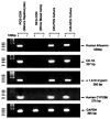
Results of RT-PCR for liver markers: PCR products were run on a 1.5% agarose gel. Lanes labeled with a (+) had reverse transcriptase added, and those with a (−) had no reverse transcriptase added. The POS-CON lane represents the positive control (mature human hepatocytes). The NEG-CON (white blood cells) represents the negative control. Both the CD45-NEG and the CD45-POS lanes, representing cultured hepatocyte-like cells derived from CD45-negative and CD45-positive SP cells, respectively, show positivity for human albumin, cytokeratin 8 (CK8), α-1 AAT (AAT), and the human cytochrome P450 gene (CYP2B6), respectively. GAPDH controls were positive for all cDNA-positive (+) samples, confirming the validity of the PCR reactions.

References

    1. Goodell MA, Brose K, Paradis G, Conner AS, Mulligan RC. Isolation and functional properties of murine hematopoietic stem cells that are replicating in vivo. J Exp Med. 1996;183:1797–1806. - PMC - PubMed
    1. Goodell MA, Rosenzweig M, Kim H, Marks DF, DeMaria M, Paradis G, Grupp SA, Sieff CA, Mulligan RC, Johnson RP. Dye efflux studies suggest that hematopoietic stem cells expressing low or undetectable levels of CD34 antigen exist in multiple species. Nat Med. 1997;3:1337–1345. - PubMed
    1. Michalopoulos GK, DeFrances MC. Liver regeneration. Science. 1997;276:60–66. - PubMed
    1. Forbes S, Vig P, Poulsom R, Thomas H, Alison M. Hepatic stem cells. J Pathol. 2002;197:510–518. - PubMed
    1. Oh SH, Hatch HM, Petersen BE. Hepatic oval ‘stem’ cell in liver regeneration. Semin Cell Dev Biol. 2002;13:405–409. - PubMed