. 2006 Mar 1;394(Pt 2):459-64.
doi: 10.1042/BJ20051123.

Semaphorin 4D activates the MAPK pathway downstream of plexin-B1

Affiliations

Semaphorin 4D activates the MAPK pathway downstream of plexin-B1

Jennifer Aurandt et al. Biochem J. .

Abstract

Semaphorins are a large family of transmembrane and secreted proteins that signal primarily through the receptor plexin. Semaphorins have been characterized in the nervous system as axon guidance cues; however, they have also been shown to control development of other cellular systems such as the vasculature and lungs. As the role of semaphorins outside of the nervous system has broadened, so has elucidation of the intracellular signalling pathways they initiate. Previously, we and others have shown that plexin-B1 activates RhoA through the binding and activation of RhoGEF (guanine nucleotide-exchange factor)/LARG (leukaemia-associated RhoGEF) in response to semaphorin 4D stimulation. In the present study, we show that semaphorin 4D activates the MAPK (mitogen-activated protein kinase) pathway. We have found that the mechanism of activation requires the C-terminus of plexin-B1 and the activation of RhoA.


Figures

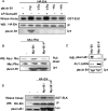
Figure 1
Figure 1. Phosphorylation of ERK in response to Sema4D treatment in HT-22 cells
(A) AP staining of HT-22 cells, a mouse hippocampal cell line. Cells were treated with specified proteins for 1 h and then stained for AP activity. (B) Presence of plexin-B1 in cell lines. Upper panel, HEK-293 (293), COS-7 and HT-22 cell lysates were analysed by Western blotting (WB) with an antibody against plexin-B1. Lower panel, characterization of antibodies against plexin-A3, -B1 and -C1. (C) Sema4D stimulates ERK phosphorylation. HT-22 cells were treated with either mock-transfected (Mock) or AP–Sema4D for 20 min. Cells were lysed and analysed by Western blotting (WB) with antibodies against phospho-ERK (pERK) and ERK. (D) Time course of ERK activation downstream of purified GST–Sema4D. HT-22 cells were treated with 4 nM GST–Sema4D for the times indicated. Cellular lysates were then analysed by Western blotting (WB) with antibodies against phospho-ERK (pERK) and ERK. (E) GST–Sema4D activates ERK. HT-22 cells were treated with 4 nM GST–Sema4D or mock-purified medium (Mock) for 20 min. Left-hand panel, cellular lysates were then analysed by Western blotting (WB) with antibodies against phospho-ERK (pERK) and ERK. Right-hand panel, Coomassie-Blue (Comm) stained GST–Sema4D and mock-purified medium (Mock).
Figure 2
Figure 2. Requirement of the C-terminus of plexin-B1 for ERK activation
(A) ERK is activated downstream of plexin-B1 in HEK-293 cells. HEK-293 cells were transfected with the indicated plasmids and then treated without (−) or with (+) AP–Sema4D. ERK kinase assay was then performed using GST–Elk as a substrate. VSV, full-length plexin-B1; -HA, C-terminally tagged full-length plexin-B1; myr, Myr–B1. Note that the level of full-length plexin-B1 transfected was below the sensitivity of the Western blot. (B) RhoA activation by Myr–B1. Rhotekin pull-down assay was performed from HEK-293 cells transfected with the indicated plasmids. Expression of Myc–Rho and Myc–B1 were determined by Western blotting (WB) of the cell lysate (Lys). Myr-B1-F, FLAG epitope tag at the C-terminus of Myr–B1. (C) AU1-LARG co-immunoprecipitates with Myr–B1. The indicated plasmids were transfected into HEK-293 cells, AU1-LARG was immunoprecipitated and a Western blot (WB) was performed with the indicated antibodies. (D) ERK activation by Myr–B1. ERK kinase assay was performed from HEK-293 cells transfected with indicated plasmids. IP, immunoprecipitation; Lys, whole-cell lysate.
Figure 3
Figure 3. Contribution of activated Rho to ERK activation
(A) Overexpression of AU1-LARG activates RhoA. A rhotekin pull-down assay was performed from HEK-293 cells transfected with the indicated plasmids. (B) ERK activation through activation of RhoA. ERK kinase assay was performed from HEK-293 cells transfected with indicated plasmids. (C) Dominant-negative LARG blocks plexin-B1 activation of ERK. ERK kinase assay was performed from HEK-293 cells transfected with Myr–B1 and increasing concentrations of the PDZ domain of LARG (Flag-DN-LARG). (D) Dominant-negative RhoA and ToxinB inhibit ERK activation. ERK kinase assay was performed from HEK-293 cells transfected with indicated plasmids. Cells were treated with ToxinB for 30 min before harvesting. (E) Dominant-negative RhoA inhibits ERK activation downstream of Sema4D/plexin-B1. HEK-293 cells were transfected with the indicated plasmids. HA–ERK was immunoprecipitated (IP) and a Western blot was performed with the indicated antibodies. Note that the level of full-length plexin-B1 transfected was below the sensitivity of the Western blot. Lys, whole-cell lysates; WB, Western blot. In panels (C) and (D), the numbers indicate the activation of ERK relative to control (arbitrary units).
Figure 4
Figure 4. Co-operation of Rho with Ras to activate the MAPK pathway
(A) Dominant-negative H-Ras inhibits activation of ERK downstream of plexin-B1. HEK-293 cells were transfected with the indicated plasmids. HA–ERK was immunoprecipitated (IP) and a Western blot (WB) was performed with the indicated antibodies. (B) Raf is activated downstream of Myr–B1. FLAG–C-Raf was co-transfected into HEK-293 cells with the indicated plasmids. FLAG–C-Raf was immunoprecipitated (IP) and a Western blot (WB) was performed with the indicated antibodies. –F, FLAG tag at the C-terminus of the protein.
Figure 5
Figure 5. Potential pathway of plexin-B1 signalling
This model represents the known signalling pathways in addition to results in the present paper. DH and PH, Dbl homology and pleckstrin homology domains respectively. MEK, MAPK/ERK kinase.

References

    1. Spriggs M. K. Shared resources between the neural and immune systems: semaphorins join the ranks. Curr. Opin. Immunol. 1999;11:387–391. - PubMed
    1. Christensen C. R., Klingelhofer J., Tarabykina S., Hulgaard E. F., Kramerov D., Lukanidin E. Transcription of a novel mouse semaphorin gene, M-semaH, correlates with the metastatic ability of mouse tumor cell lines. Cancer Res. 1998;58:1238–1244. - PubMed
    1. Behar O., Golden J. A., Mashimo H., Schoen F. J., Fishman M. C. Semaphorin III is needed for normal patterning and growth of nerves, bones and heart. Nature (London) 1996;383:525–528. - PubMed
    1. Basile J. R., Barac A., Zhu T., Guan K. L., Gutkind J. S. Class IV semaphorins promote angiogenesis by stimulating Rho-initiated pathways through plexin-B. Cancer Res. 2004;64:5212–5224. - PubMed
    1. Ito T., Kagoshima M., Sasaki Y., Li C., Udaka N., Kitsukawa T., Fujisawa H., Taniguchi M., Yagi T., Kitamura H., Goshima Y. Repulsive axon guidance molecule Sema3A inhibits branching morphogenesis of fetal mouse lung. Mech. Dev. 2000;97:35–45. - PubMed

Publication types

MeSH terms