. 2005 Oct 15;118(Pt 20):4709-20.
doi: 10.1242/jcs.02598. Epub 2005 Sep 27.

Receptor-activity-modifying proteins are required for forward trafficking of the calcium-sensing receptor to the plasma membrane

Affiliations

Receptor-activity-modifying proteins are required for forward trafficking of the calcium-sensing receptor to the plasma membrane

Tristan Bouschet et al. J Cell Sci. .

Abstract

The calcium-sensing receptor (CaSR) is a class III G-protein-coupled receptor (GPCR) that responds to changes in extracellular calcium concentration and plays a crucial role in calcium homeostasis. The mechanisms controlling CaSR trafficking and surface expression are largely unknown. Using a CaSR tagged with the pH-sensitive GFP super-ecliptic pHluorin (SEP-CaSR), we show that delivery of the GPCR to the cell surface is dependent on receptor-activity-modifying proteins (RAMPs). We demonstrate that SEP-CaSRs are retained in the endoplasmic reticulum (ER) in COS7 cells that do not contain endogenous RAMPs whereas they are delivered to the plasma membrane in HEK 293 cells that do express RAMP1. Coexpression of RAMP1 or RAMP3, but not RAMP2, in COS7 cells was sufficient to target the CaSR to the cell surface. RAMP1 and RAMP3 colocalised and coimmunoprecipitated with the CaSR suggesting that these proteins associate within the cell. Our results indicate that RAMP expression promotes the forward trafficking of the GPCR from the ER to the Golgi apparatus and results in mature CaSR glycosylation, which is not observed in RAMP-deficient cells. Finally, silencing of RAMP1 in the endogenously expressing HEK293 cells using siRNA resulted in altered CaSR traffic. Taken together, our results show that the association with RAMPs is necessary and sufficient to transfer the immature CaSR retained in the ER towards the Golgi where it becomes fully glycosylated prior to delivery to the plasma membrane and demonstrate a role for RAMPs in the trafficking of a class III GPCR.


Figures

Fig. 1
Fig. 1
Functional characterisation of SEP-CaSR. (A) Schematic representation of super-ecliptic pHluorin-tagged calcium-sensing receptor (SEP-CaSR). The plasma membrane and the flytrap domain are indicated. (B) Live confocal imaging of typical SEP-CaSR fluorescence distribution in two cultured HEK cells transfected with SEP-CaSR. Note that fluorescence is mainly visible at the plasma membrane. SEP-CaSR surface expression was assessed by monitoring fluorescence intensity in response to pH changes: reducing extracellular pH from pH 7.4 (Control, Ctl) to pH 6 causes a decrease in fluorescence as surface SEP-CaSR is eclipsed. Surface fluorescence is restored following a rinse with the pH 7.4 control solution. Application of a solution at pH 7.4 containing NH4Cl (50 mM), which rapidly equilibrates cellular pH levels, causes a sharp increase as all the SEP-CaSR fluorescence in the cell is revealed. The intracellular fluorescence disappears following a rinse with the control solution. (C) Immunoblot of SEP-CaSR. Cell extracts (~20 μg) from HEK cells expressing pRK5 (empty vector, as a control), pRK5-CaSR or pRK5-SEP-CaSR were run on an 8% polyacrylamide gel and probed with anti-CaSR ADD antibody (left panel) or with anti-GFP antibody (right panel). Position of molecular mass markers is indicated and arrowheads indicate the putative monomeric and homodimeric forms of SEP-CaSR. (D) Intracellular calcium concentration is increased by SEP-CaSR activation. HEK293 cells were transfected with SEP-CaSR (green) and loaded with the calcium-sensitive dye Fura Red (red). Merged areas appear yellow. The corresponding transmitted light image is shown. (E) Graph of the time courses of fluorescence from the regions selected in the fluorescence and transmitted light images in D. Stimulation with 5 mM CaCl2 causes oscillations in the Fura Red fluorescence intensity, which corresponds to [Ca2+]i oscillations, in the SEP-CaSR-expressing cell. The data are representative of three independent experiments. Bar, 10 μm.
Fig. 2
Fig. 2
Cell-type-specific CaSR surface expression. (A) SEP-CaSR expression at the plasma membrane. Surface CaSR was labelled without permeabilisation in HEK293 cells expressing SEP-CaSR using a rabbit anti-GFP antibody followed by a Cy3 anti-rabbit antibody. Colocalisation of the SEP-CaSR fluorescence (green) and surface-labelled SEP-CaSR (Cy3, red) is revealed in yellow. A corresponding transmitted light image is shown to highlight the localisation of SEP-CaSR at the plasma membrane (arrowheads). Representative micrographs of five independent experiments are shown. (B) Confocal images showing differential CaSR distribution in HEK293 and COS7 cells expressing SEP-CaSR. Surface CaSR was labelled without permeabilisation using a rabbit anti-GFP antibody followed by a Cy3 anti-rabbit antibody. The overlay of total (green, left) and surface SEP-CaSR (Cy3, red, centre) is revealed in yellow (right). Arrowheads indicate the plasma membrane. A corresponding transmitted light image is shown for COS7 cells to highlight the lack of staining at the plasma membrane. Representative micrographs of three independent experiments are shown. (C) Differential CaSR cell surface expression assessed by surface biotinylation experiments. Surface proteins from COS7 and HEK293 cells transfected with SEP-CaSR were isolated by surface biotinylation as described in the Methods and the total and surface CaSR populations revealed by western blotting using an antibody against GFP. Representative blots of four independent experiments are shown. (D) Cell-type-restricted RAMP mRNA expression. RT-PCR amplifications of RAMP1, RAMP2 and RAMP3 mRNA were performed on samples from HEK293 and COS7 cell cultures and resolved on an agarose gel. A similar expression pattern was obtained in all the preparations tested (three independent experiments). Before PCR amplification, samples were (+) or not (−) submitted to reverse transcriptase (RT) treatment to estimate DNA contamination. GAPDH was used as an internal control and sizes of expected products are indicated. Bar, 5 μm (A); 10 μm (B).
Fig. 3
Fig. 3
RAMP1 and RAMP3, but not RAMP2, promote cell surface delivery of the CaSR. (A) Effect of RAMPs on CaSR surface expression assessed by surface biotinylation. COS7 cells were transfected with SEP-CaSR in combination with a control vector (encoding CAT) or vectors encoding RAMP1, RAMP2 or RAMP3. 48 hours post-transfection, the total and cell surface CaSR populations were determined as described in Fig. 2 legend and Materials and Methods. A representative western blot of four independent experiments is shown. (B) Densitometric analysis of the effect of RAMPs on CaSR surface expression measured by biotinylation assays as illustrated in A. Results are the mean±s.e.m. of four independent experiments. *P≤0.001 compared with levels in the control (Student’s t-test). (C) Effect of RAMPs on CaSR surface expression assessed by immunofluorescence. COS7 were transfected with plasmids encoding SEP-CaSR and Myc-RAMP1, HA-RAMP2, HA-RAMP3 or CAT (as a control). Cells were then stained for surface CaSR using sequentially rabbit anti-GFP and Cy3 anti-rabbit antibodies. Cells were then permeabilised and Myc-RAMP1, HA-RAMP2 or HA-RAMP3 were labelled using mouse anti-Myc or anti-HA antibodies followed by Cy5 anti-mouse antibodies. Surface CaSR and RAMPs appear in red and blue respectively. A transmitted light image is shown for the control condition. Images are representative of three independent experiments. Bar, 10 μm.
Fig. 4
Fig. 4
Internal and surface colocalisation experiments of CaSR with RAMPs. (A) COS7 were transfected with plasmids encoding SEP-CaSR and Myc-RAMP1, HA-RAMP2, HA-RAMP3 or CAT (as a control). 48 hours post-transfection, cells were stained in permeabilised conditions (see methods) for CaSR and RAMPs using respectively rabbit anti-GFP and mouse anti-HA or Myc antibodies followed by Cy5 anti-rabbit or Cy3 anti-mouse antibodies. Colocalisation regions of RAMPs (red) and CaSR (blue) appear in purple and are indicated by arrowheads. Images are representative of three independent experiments. (B) Surface colocalisation of CaSR and RAMP3. COS7 cells were transfected with either control plasmid (upper panels) or SEP-CaSR in the absence (middle panels) or presence (lower panels) of HA-RAMP3. Cells were incubated in non-permeabilised conditions with anti-GFP and anti-HA antibodies followed by Cy5 anti-rabbit and Cy3 anti-mouse antibodies to label surface CaSR and surface RAMP3 respectively (see Materials and Methods). Regions of colocalisation between surface CaSR (blue) and surface RAMP (red) appear purple and triple colocalisation areas (white) are indicated by arrowheads. Images are representative of three independent experiments. Bar, 10 μm.
Fig. 5
Fig. 5
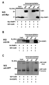
The CaSR associates with RAMP1 and RAMP3. COS7 cells were transfected with SEP-CaSR in combination with Myc-RAMP1 and HA-RAMP3 as indicated. 48 hours post-transfection, cells were lysed and 600 μg of proteins were immunoprecipitated with monoclonal anti-HA, anti-Myc or anti-GFP antibodies. (A) RAMP1 is present in CaSR immunoprecipitates. Western blot (W.B.) with anti-Myc antibody (to detect Myc-RAMP1) of total proteins (input, lanes 1 and 2) and of immunoprecipitates isolated by incubation with indicated antibodies (lanes 3-6). Myc-RAMP1 was retrieved in anti-GFP immunoprecipitates prepared from COS7 cells expressing both the SEP-CaSR and Myc-RAMP1 (lane 3) but not when the anti-GFP antibody or the SEP-CaSR coding plasmid were omitted (lanes 4 and 5 respectively), indicating that the binding of Myc-RAMP1 occurs in a CaSR-dependent manner. Immunoprecipitation using anti-Myc antibodies indicates the expected size of immunoprecipitated Myc-RAMP1 (lane 6). (B) RAMP3 is present in CaSR immunoprecipitates. Immunoblot using anti-HA antibody (to detect HA-RAMP3) of total proteins (input, lanes 1 and 2) and of immunoprecipitates (lanes 3-5). RAMP3 was retrieved in anti-GFP immunoprecipitates prepared from COS7 cells expressing both the SEP-CaSR and HA-RAMP3 (lane 3) but not when the SEP-CaSR plasmid was omitted (lane 4), indicating that the binding of the HA-RAMP3 occurs in a CaSR-dependent manner. A positive control immunoprecipitate using anti-HA antibodies was loaded to indicate the expected size of HA-RAMP3 (lane 5). (C) The CaSR is present in RAMP3 precipitates. Western blot using anti-GFP antibody (to detect SEP-CaSR) of total proteins (input, lanes 1 and 2) and immunoprecipitates (lanes 3-5). The CaSR was retrieved in anti-HA immunoprecipitates prepared from COS7 cells expressing both the SEP-CaSR and HA-RAMP3 (lane 3) but not when HA-RAMP3 plasmid was omitted (lane 4), indicating that the binding of the CaSR occurs in a RAMP3-dependent manner. Immunoprecipitation using anti-GFP antibodies indicates the expected size of immunoprecipitated SEP-CaSR (lane 5). The monomeric and dimeric SEP-CaSR forms are indicated.
Fig. 6
Fig. 6
RAMP stimulates the traffic of the CaSR from the ER to the Golgi apparatus. Subcellular localisation of the SEP-CaSR in the ER and the Golgi apparatus in COS7 cells expressing the receptor in the absence (upper panels, a-h) or in the presence of RAMP (HA-RAMP3, lower panels, i-o). Cells were fixed and the ER and the Golgi apparatus were stained with antibodies directed against protein markers calnexin and giantin, respectively. In parallel, cells were stained with HA antibody to label HA-RAMP3 and a final wash with a solution at pH of 7.4 containing 50 mM NH4Cl was performed in order to illuminate the entire SEP-CaSR population (as shown in Fig. 1B). The colors expected for merged areas are indicated. Images are representative of three independent experiments. Bar, 20 μm.
Fig. 7
Fig. 7
RAMPs promote terminal glycosylation of the CaSR. Proteins (30 μg) from HEK293 and COS7 cells expressing SEP-CaSR in the absence or presence of HA-RAMP3 were deglycosylated by incubation with Endo-H or PNGase-F. The samples were subsequently resolved by SDS-PAGE and the different states of glycosylation of the CaSR were revealed by probing with an anti-GFP antibody. The presence or absence of enzyme treatment are respectively indicated by the + and − signs. Gels shown are representative of three independent experiments.
Fig. 8
Fig. 8
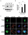
Depletion of RAMP1 in HEK293 cells alters CaSR traffic. (A) Silencing of RAMP1 using siRNA. HEK293 cells were transfected with 20 nM siRNAs (control, siRAMP1# 1, siRAMP1 #2, see Materials and Methods) and 500 ng Myc-RAMP1. Whole cell lysates were prepared 48 hours after transfection. The immunoblot was probed with anti-Myc antibody and reprobed with anti-β-actin antibody to ensure equal loading. Films were scanned and quantified using the image J software. A significant decrease of RAMP1 expression (*P<0.05) was observed with siRAMP1 #2 compared to the expression level in the control. The blots shown are representative of three independent experiments. (B) Silencing RAMP1 alters CaSR traffic. HEK293 cells were transfected with 20 nM Cy3-labelled siRNA against RAMP1 (siRAMP1 #1 and #2) or control siRNAs together with 200 ng pf pRK5-SEP-CaSR. 48 hours after transfection, the surface expressed SEP-CaSR population was labelled with Alexa 655 anti-GFP antibody, cells were fixed and imaging was performed as described in Materials and Methods. SEP-CaSR appears green, Cy3-siRNAs red and surface CaSR blue. Bar, 5 μm.
Fig. 9
Fig. 9
Proposed model for RAMP regulation of CaSR trafficking. (A) In the absence of RAMPs (COS7 cells), the CaSR is retained (stop sign) in the endoplasmic reticulum (calnexin-positive compartment) in its incompletely processed, core-glycosylated form. (B) By contrast, in cells expressing RAMP endogenously (such as HEK293 cells), or exogenously (such as RAMP-transfected COS7 cells), the CaSR in association with RAMP1 or RAMP3 bypasses the ER retention and reaches the Golgi apparatus (giantin-positive compartment) where it is terminally glycosylated before being delivered to the cell surface.

Similar articles

Cited by

References

    1. Ashby MC, De La Rue SA, Ralph GS, Uney J, Collingridge GL, Henley JM. Removal of AMPA receptors (AMPARs) from synapses is preceded by transient endocytosis of extrasynaptic AMPARs. J. Neurosci. 2004a;24:5172–5176. - PMC - PubMed
    1. Ashby MC, Ibaraki K, Henley JM. It’s green outside: tracking cell surface proteins with pH-sensitive GFP. Trends Neurosci. 2004b;27:257–261. - PubMed
    1. Awata H, Huang C, Handlogten ME, Miller RT. Interaction of the calcium-sensing receptor and filamin, a potential scaffolding protein. J. Biol. Chem. 2001;276:34871–34879. - PubMed
    1. Bai M, Trivedi S, Brown EM. Dimerization of the extracellular calcium-sensing receptor (CaR) on the cell surface of CaR-transfected HEK293 cells. J. Biol. Chem. 1998;273:23605–23610. - PubMed
    1. Berridge MJ, Lipp P, Bootman MD. The versatility and universality of calcium signalling. Nat. Rev. Mol. Cell Biol. 2000;1:11–21. - PubMed

Publication types

MeSH terms

Substances