L-domain flanking sequences are important for host interactions and efficient budding of vesicular stomatitis virus recombinants
- PMID: 16188963
- PMCID: PMC1235845
- DOI: 10.1128/JVI.79.20.12617-12622.2005
L-domain flanking sequences are important for host interactions and efficient budding of vesicular stomatitis virus recombinants
Abstract
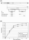
Vesicular stomatitis virus (VSV) possesses a PPPY and a PSAP motif within the matrix (M) protein. The PPPY motif has significant L-domain activity in BHK-21 cells, whereas the PSAP motif does not. Since the core PSAP motif alone is insufficient to provide L-domain activity, we modified upstream or downstream amino acids flanking the PSAP core motif to determine their effect on L-domain activity. VSV recombinants were recovered that contained single or multiple amino acid mutations in upstream or downstream sequences flanking the PSAP core. Recombinant viruses were examined for growth kinetics, budding efficiency, and functional interactions with host proteins. We demonstrate that the composition of amino acids surrounding the L-domain core motifs are critical for efficient L-domain activity and for interactions with host proteins in the context of a VSV infection.
Figures


Similar articles
-
Functional analysis of late-budding domain activity associated with the PSAP motif within the vesicular stomatitis virus M protein.J Virol. 2004 Jul;78(14):7823-7. doi: 10.1128/JVI.78.14.7823-7827.2004. J Virol. 2004. PMID: 15220457 Free PMC article.
-
Modifications of the PSAP region of the matrix protein lead to attenuation of vesicular stomatitis virus in vitro and in vivo.J Gen Virol. 2007 Sep;88(Pt 9):2559-2567. doi: 10.1099/vir.0.83096-0. J Gen Virol. 2007. PMID: 17698667
-
Phenotypes of vesicular stomatitis virus mutants with mutations in the PSAP motif of the matrix protein.J Gen Virol. 2012 Apr;93(Pt 4):857-865. doi: 10.1099/vir.0.039800-0. Epub 2011 Dec 21. J Gen Virol. 2012. PMID: 22190013
-
Vesicular stomatitis virus as an oncolytic vector.Viral Immunol. 2004;17(4):516-27. doi: 10.1089/vim.2004.17.516. Viral Immunol. 2004. PMID: 15671748 Review.
-
Contributions of vesicular stomatitis virus to the understanding of RNA virus evolution.Curr Opin Microbiol. 2003 Aug;6(4):399-405. doi: 10.1016/s1369-5274(03)00084-5. Curr Opin Microbiol. 2003. PMID: 12941412 Review.
Cited by
-
Exosomal transmission of viruses, a two-edged biological sword.Cell Commun Signal. 2023 Jan 23;21(1):19. doi: 10.1186/s12964-022-01037-5. Cell Commun Signal. 2023. PMID: 36691072 Free PMC article. Review.
-
PPEY motif within the rabies virus (RV) matrix protein is essential for efficient virion release and RV pathogenicity.J Virol. 2008 Oct;82(19):9730-8. doi: 10.1128/JVI.00889-08. Epub 2008 Jul 30. J Virol. 2008. PMID: 18667490 Free PMC article.
-
Cytopathogenesis of vesicular stomatitis virus is regulated by the PSAP motif of M protein in a species-dependent manner.Viruses. 2012 Sep;4(9):1605-18. doi: 10.3390/v4091605. Epub 2012 Sep 19. Viruses. 2012. PMID: 23170175 Free PMC article.
-
Mechanisms for enveloped virus budding: can some viruses do without an ESCRT?Virology. 2008 Mar 15;372(2):221-32. doi: 10.1016/j.virol.2007.11.008. Epub 2007 Dec 11. Virology. 2008. PMID: 18063004 Free PMC article. Review.
-
Ilaprazole and other novel prazole-based compounds that bind Tsg101 inhibit viral budding of HSV-1/2 and HIV from cells.J Virol. 2021 May 10;95(11):e00190-21. doi: 10.1128/JVI.00190-21. Epub 2021 Mar 17. J Virol. 2021. PMID: 33731460 Free PMC article.
References
-
- Blot, V., F. Perugi, B. Gay, M. C. Prevost, L. Briant, F. Tangy, H. Abriel, O. Staub, M. C. Dokhelar, and C. Pique. 2004. Nedd4.1-mediated ubiquitination and subsequent recruitment of Tsg101 ensure HTLV-1 Gag trafficking towards the multivesicular body pathway prior to virus budding. J. Cell Sci. 117:2357-2367. - PubMed
-
- Bouamr, F., J. A. Melillo, M. Q. Wang, K. Nagashima, M. de Los Santos, A. Rein, and S. P. Goff. 2003. PPPYVEPTAP motif is the late domain of human T-cell leukemia virus type 1 Gag and mediates its functional interaction with cellular proteins Nedd4 and Tsg101. J. Virol. 77:11882-11895. - PMC - PubMed
Publication types
MeSH terms
Substances
Grants and funding
LinkOut - more resources
Full Text Sources
Other Literature Sources
Research Materials
Miscellaneous

