. 2005 Oct;79(20):12721-31.
doi: 10.1128/JVI.79.20.12721-12731.2005.

ADP-ribose-1"-monophosphatase: a conserved coronavirus enzyme that is dispensable for viral replication in tissue culture

Affiliations

ADP-ribose-1"-monophosphatase: a conserved coronavirus enzyme that is dispensable for viral replication in tissue culture

Akos Putics et al. J Virol. 2005 Oct.

Abstract

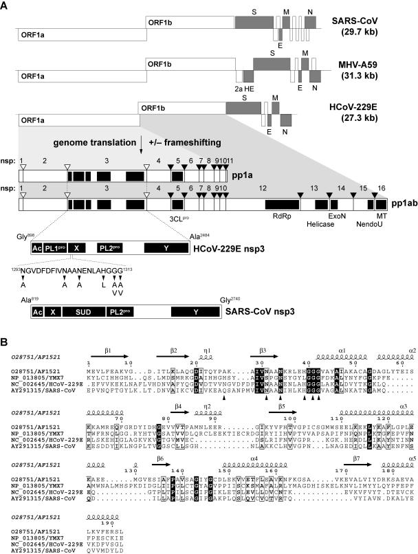
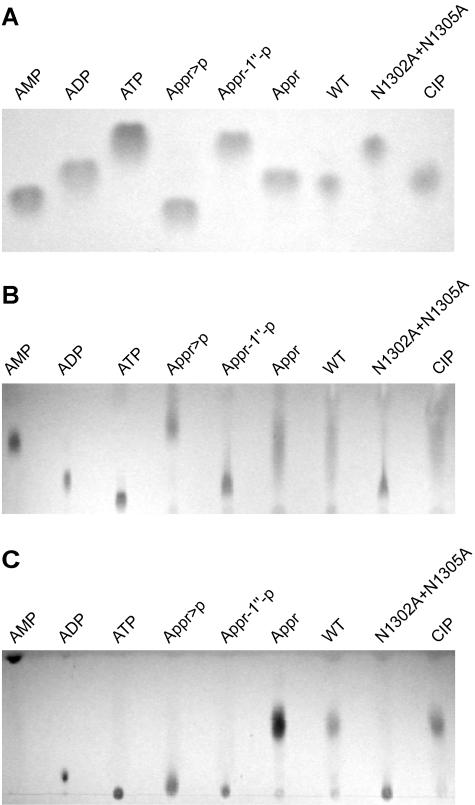
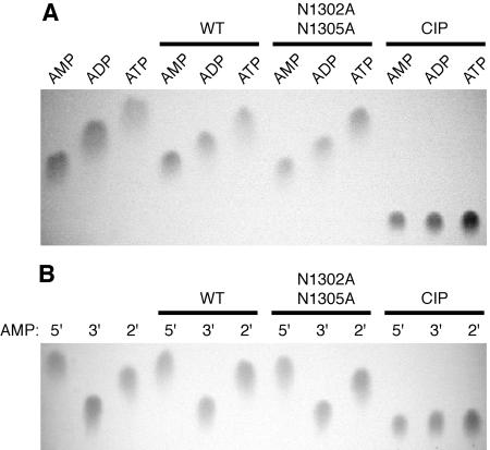
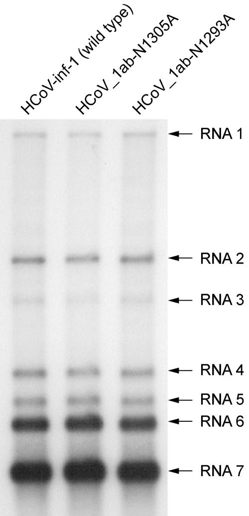
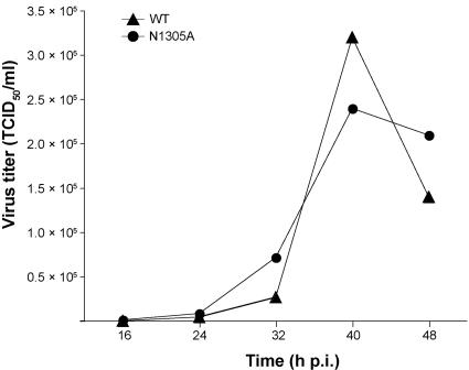
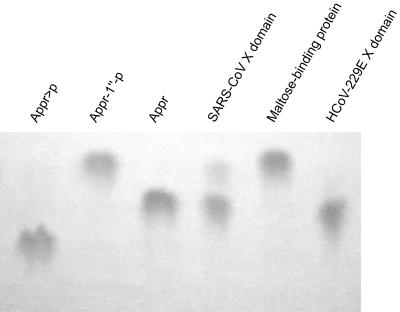
Replication of the approximately 30-kb plus-strand RNA genome of coronaviruses and synthesis of an extensive set of subgenome-length RNAs are mediated by the replicase-transcriptase, a membrane-bound protein complex containing several cellular proteins and up to 16 viral nonstructural proteins (nsps) with multiple enzymatic activities, including protease, polymerase, helicase, methyltransferase, and RNase activities. To get further insight into the replicase gene-encoded functions, we characterized the coronavirus X domain, which is part of nsp3 and has been predicted to be an ADP-ribose-1"-monophosphate (Appr-1"-p) processing enzyme. Bacterially expressed forms of human coronavirus 229E (HCoV-229E) and severe acute respiratory syndrome-coronavirus X domains were shown to dephosphorylate Appr-1"-p, a side product of cellular tRNA splicing, to ADP-ribose in a highly specific manner. The enzyme had no detectable activity on several other nucleoside phosphates. Guided by the crystal structure of AF1521, an X domain homolog from Archaeoglobus fulgidus, potential active-site residues of the HCoV-229E X domain were targeted by site-directed mutagenesis. The data suggest that the HCoV-229E replicase polyprotein residues, Asn 1302, Asn 1305, His 1310, Gly 1312, and Gly 1313, are part of the enzyme's active site. Characterization of an Appr-1"-pase-deficient HCoV-229E mutant revealed no significant effects on viral RNA synthesis and virus titer, and no reversion to the wild-type sequence was observed when the mutant virus was passaged in cell culture. The apparent dispensability of the conserved X domain activity in vitro indicates that coronavirus replicase polyproteins have evolved to include nonessential functions. The biological significance of the novel enzymatic activity in vivo remains to be investigated.


Figures

FIG. 1.
FIG. 1.
Coronavirus replicase gene-encoded X domains reside in nonstructural protein 3. (A) Structures of the 5′ capped and 3′ adenylated plus-strand RNA genomes of SARS-CoV, MHV-A59, and HCoV-229E are shown. Functional ORFs in the genome are expressed from the genomic RNA and an extensive set of subgenomic mRNAs. ORFs encoding the coronavirus structural proteins, that is, the spike (S), envelope (E), membrane (M), nucleocapsid (N), and (in MHV-A59) hemagglutinin-esterase (HE) proteins are indicated in gray. MHV-A59 ORF2a has been predicted to encode a cyclic phosphodiesterase activity that is not conserved in SARS-CoV and HCoV-229E (48). The 5′ terminal replicase gene, which is comprised of ORF1a and ORF1b, encodes the replicative polyproteins pp1a and, by −1 ribosomal frameshifting, a C-terminally extended version of pp1a, which is called pp1ab. The two polyproteins are cleaved by viral proteases to yield the 16 processing end products nsps 1 to 16. Cleavage sites processed by the papain-like proteases, PL1pro and PL2pro, are indicated by open triangles, and cleavage sites processed by the 3C-like main protease, 3CLpro, are indicated by filled triangles. Conserved coronavirus replicase gene-encoded domains are illustrated for the HCoV-229E pp1a and pp1ab (48, 59). Details on the nsp3 subdomain organizations of HCoV-229E and SARS-CoV, whose X domains were characterized in this study, are given below. Please note that SARS-CoV nsp3 lacks a counterpart of PL1pro and encodes an extra domain downstream of X, the SARS-CoV unique domain, which is not conserved in other coronaviruses. The HCoV-229E X domain single-residue substitutions characterized in this study are indicated. RdRp, RNA-dependent RNA polymerase; ExoN, putative 3′-to-5′ exoribonuclease; NendoU, nidoviral uridylate-specific endoribonuclease (23); MT, putative ribose-2′-O methyltransferase; Ac, acidic domain; Y, domain containing conserved Cys and His residues and stretches of hydrophobic residues presumed to anchor nsp3 to intracellular membranes (63). (B) Sequence comparison of the X domains of HCoV-229E and SARS-CoV with cellular homologs from A. fulgidus (AF1521) (1) and S. cerevisiae (YMX7) (27). The presented set was derived from an alignment of dozens of virus and cellular homologs that was generated using the ClustalX program (versions 1.64 and 1.8) (6) with manual adjustment and cross-verification with an alignment of coronavirus nsp3 proteins (63) (A. E. Gorbalenya and J. Ziebuhr, unpublished data). The secondary structure information for AF1521 was derived from the published crystal structure (1) (Protein Data Bank accession no. 1VHU) and, together with the alignment, was used as input for the ESPript program, version 2.2 (http://prodes.toulouse.inra.fr/ESPript/cgi-bin/ESPript.cgi). Sequences of the proteins were derived from the DDBJ/EMBL/GenBank database accession numbers O28751 (for AF1521), NP_013805 (for YMX7), NC_002645 (HCoV-229E X domain, pp1a/pp1ab residues Glu 1265 to Leu 1435), and AY291315 (SARS-CoV X domain, pp1a/pp1ab residues Glu 1000 to Asp 1170). The positions of HCoV-229E X domain residues that were substituted in this study are indicated by arrowheads (see also panel A).
FIG. 2.
FIG. 2.
Bacterially expressed HCoV-229E X domain converts Appr-1"-p to Appr. (A) The HCoV-229E X domain (pp1a/pp1ab residues 1265 to 1436) was expressed and purified as an MBP fusion protein (see Materials and Methods) and subsequently cleaved with factor Xa. This protein and a mutant derivative carrying two substitutions of conserved Asn residues were incubated for 3 h at 30°C with Appr-1"-p that was generated from Appr>p using A. thaliana CPDase (see Materials and Methods). Lanes: WT, incubation with HCoV-229E X (wild-type sequence); N1302A+N1305A, incubation with a mutant form of HCoV-229E X in which the pp1a/pp1ab Asn residues 1302 and 1305 were each substituted with Ala; CIP, incubation with alkaline phosphatase isolated from calf intestine. Reaction products were separated on cellulose TLC plates using solvent I and visualized under UV light. Marker nucleotides: AMP, ADP, ATP, Appr, Appr-1"-p, and Appr>p. (B and C) The reactions described in panel A were analyzed on polyethyleneimine-cellulose TLC plates using solvent II (B) and III (C), respectively.
FIG. 3.
FIG. 3.
The phosphatase activity of the HCoV-229E X domain is highly specific for Appr-1"-p. (A) The adenosine phosphates, AMP, ADP, and ATP, respectively, were incubated for 3 h at 30°C with the following proteins: WT, HCoV-229E X (wild-type sequence); N1302A+N1305A, mutant form of HCoV-229E X in which the pp1a/pp1ab Asn residues 1302 and 1305 were each replaced by Ala; CIP, alkaline phosphatase isolated from calf intestine. (B) The adenosine monophosphates, 5′ AMP (5′),3′ AMP (3′), and 2′ AMP (2′), respectively, were incubated with HCoV-229E X (WT; wild-type sequence),a mutant form of HCoV-229E X (N1302A+N1305A), and CIP. In both experiments, the reaction products were separated on cellulose TLC plates using solvent I as liquid phase. Nucleotide markers: AMP, ADP, ATP, 5′ AMP, 3′ AMP, and 2′ AMP.
FIG. 4.
FIG. 4.
Activities of mutant forms of the HCoV-229E Appr-1"-pase. (A and B) Coomassie brilliant blue-stained sodium dodecyl sulfate-polyacrylamide gels showing the bacterially expressed HCoV-229E X domain (wild-type sequence) and the mutant forms characterized in this study (substitutions indicated) after purification and factor Xa-mediated release from the MBP fusion protein. (C and D) Appr-1"-pase activities of HCoV-229E Appr-1"-pases containing the indicated substitutions of conserved and nonconserved residues (Fig. 1 and Table 2). Incubation of the substrate Appr-1"-p with HCoV-229E Appr-1"-pase (wild-type sequence [WT]) served as a positive control, and for each of the reactions an equal amount of purified protein was used (for details, see Material and Methods). Reactions were performed at 30°C for 3 h and the reaction products were analyzed by cellulose TLC using solvent I as liquid phase. The positions of nucleotide markers (Appr-1"-p and Appr) are indicated by arrowheads.
FIG. 5.
FIG. 5.
RNA synthesis of the HCoV-229E Appr-1"-pase mutants HCoV_1ab-N1293A and HCoV_1ab-N1305. Shown is a Northern blot analysis of viral RNA synthesis at 72 h postinfection using poly(A) RNA from 6 × 10⁵ MRC-5 cells that had been infected with HCoV-229E (wild type [WT]), HCoV_1ab-N1293A, and HCoV_1ab-N1305A, respectively, at an MOI of 0.01 TCID50 per cell. The 32P-labeled probe used in this experiment was specific for the HCoV-229E 3′ terminal nucleotides 26857 to 27235.
FIG. 6.
FIG. 6.
Growth kinetics in MRC-5 cells of an HCoV-229E mutant with an Appr-1"-pase active-site substitution. Virus titers (given as mean values of two independent experiments) of wild-type HCoV-229E (WT) and HCoV_1ab-N1305A (N1305A) were determined at the indicated times points as described in Materials and Methods. p.i., postinfection.
FIG. 7.
FIG. 7.
Appr-1"-pase activity of the SARS-CoV X domain. The X domain of SARS-CoV (pp1a/pp1ab residues 1000 to 1173) was overexpressed as an E. coli MBP fusion protein, purified by amylose-affinity chromatography and released from MBP by factor Xa treatment. Following incubation of the Appr-1"-p substrate with the SARS-CoV X domain for 3 h at 30°C, the reaction products were separated by cellulose TLC using solvent I and visualized under UV light. As positive and negative controls, respectively, the HCoV-229E X domain and MBP were used. Appr-1"-p, Appr, and Appr>p were used as markers.

References

    1. Allen, M. D., A. M. Buckle, S. C. Cordell, J. Lowe, and M. Bycroft. 2003. The crystal structure of AF1521 a protein from Archaeoglobus fulgidus with homology to the non-histone domain of macroH2A. J. Mol. Biol. 330:503-511. - PubMed
    1. Anand, K., G. J. Palm, J. R. Mesters, S. G. Siddell, J. Ziebuhr, and R. Hilgenfeld. 2002. Structure of coronavirus main proteinase reveals combination of a chymotrypsin fold with an extra alpha-helical domain. EMBO J. 21:3213-3224. - PMC - PubMed
    1. Anand, K., J. Ziebuhr, P. Wadhwani, J. R. Mesters, and R. Hilgenfeld. 2003. Coronavirus main proteinase (3CLpro) structure: basis for design of anti-SARS drugs. Science 300:1763-1767. - PubMed
    1. Bhardwaj, K., L. Guarino, and C. C. Kao. 2004. The severe acute respiratory syndrome coronavirus Nsp15 protein is an endoribonuclease that prefers manganese as a cofactor. J. Virol. 78:12218-12224. - PMC - PubMed
    1. Brierley, I., M. E. Boursnell, M. M. Binns, B. Bilimoria, V. C. Blok, T. D. Brown, and S. C. Inglis. 1987. An efficient ribosomal frame-shifting signal in the polymerase-encoding region of the coronavirus IBV. EMBO J. 6:3779-3785. - PMC - PubMed

Publication types

MeSH terms

LinkOut - more resources