. 2005 Oct;79(20):12783-97.
doi: 10.1128/JVI.79.20.12783-12797.2005.

Site-directed mutagenesis of large DNA palindromes: construction and in vitro characterization of herpes simplex virus type 1 mutants containing point mutations that eliminate the oriL or oriS initiation function

Affiliations

Site-directed mutagenesis of large DNA palindromes: construction and in vitro characterization of herpes simplex virus type 1 mutants containing point mutations that eliminate the oriL or oriS initiation function

John W Balliet et al. J Virol. 2005 Oct.

Abstract

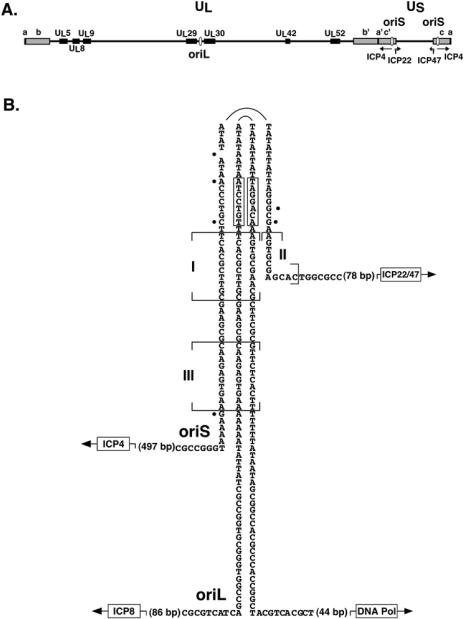
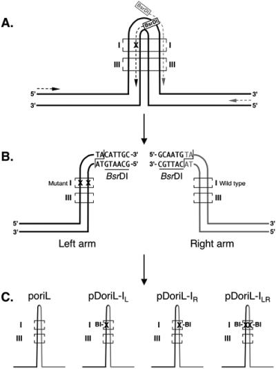
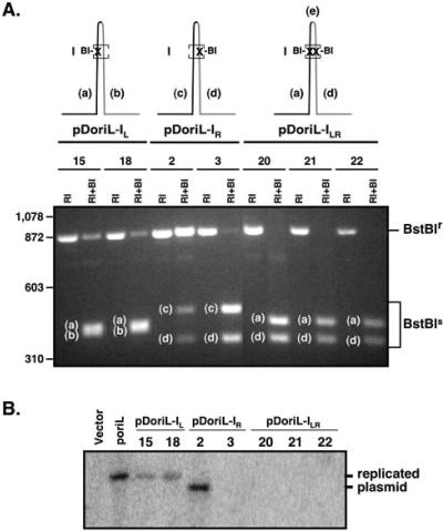
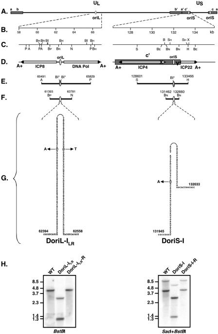
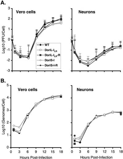
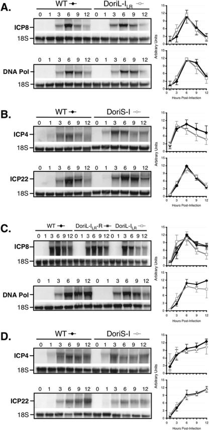
Technical challenges associated with mutagenesis of the large oriL palindrome have hindered comparisons of the functional roles of the herpes simplex virus type 1 (HSV-1) origins of DNA replication, oriL and oriS, in viral replication and pathogenesis. To address this problem, we have developed a novel PCR-based strategy to introduce site-specific mutations into oriL and other large palindromes. Using this strategy, we generated three plasmids containing mutant forms of oriL, i.e., pDoriL-I(L), pDoriL-I(R), and pDoriL-I(LR), containing point mutations in the left, right, and both copies, respectively, of the origin binding protein (OBP) binding site (site I) which eliminate OBP binding. In in vitro DNA replication assays, plasmids with mutations in only one arm of the palindrome supported origin-dependent DNA replication, whereas plasmids with symmetrical mutations in both arms of the palindrome were replication incompetent. An analysis of the cloned mutant plasmids used in replication assays revealed that a fraction of each plasmid mutated in only one arm of the palindrome had lost the site I mutation. In contrast, plasmids containing symmetrical mutations in both copies of site I retained both mutations. These observations demonstrate that the single site I mutations in pDoriL-I(L) and pDoriL-I(R) are unstable upon propagation in bacteria and suggest that functional forms of both the left and right copies of site I are required to initiate DNA replication at oriL. To examine the role of oriL and oriS site I in virus replication, we introduced the two site I mutations in pDoriL-I(LR) into HSV-1 DNA to yield the mutant virus DoriL-I(LR) and the same point mutations into the single site I sequence present in both copies of oriS to yield the mutant virus DoriS-I. In Vero cells and primary rat embryonic cortical neurons (PRN) infected with either mutant virus, viral DNA synthesis and viral replication were efficient, confirming that the two origins can substitute functionally for one another in vitro. Measurement of the levels of oriL and oriS flanking gene transcripts revealed a modest alteration in the kinetics of ICP8 transcript accumulation in DoriL-I(LR)-infected PRN, but not in Vero cells, implicating a cell-type-specific role for oriL in regulating ICP8 transcription.


Figures

FIG. 1.
FIG. 1.
Genomic locations and sequence alignment of HSV-1 oriL and oriS. (A) Diagram of the HSV-1 genome showing the UL region flanked by inverted repeat sequences, ab and b′a′, and the US region flanked by inverted repeat sequences, a′c′ and ca. The locations of oriL and both copies of oriS are indicated by white ovals. Black boxes indicate the locations of the seven E genes that are necessary and sufficient for viral origin-dependent DNA replication (41). Two essential E genes, UL29 and UL30, are divergently transcribed from oriL and encode the single-stranded DNA binding protein (ICP8) and Pol, respectively. Three IE genes encoding viral regulatory proteins ICP4, -22, and -47 are divergently transcribed from oriS; these transcripts are shown as black arrows. (B) Alignment of nucleotide sequences in the oriL and oriS palindromes. For simplicity and to better illustrate regions of sequence homology, the nucleotide sequences of both origins are presented as palindromes, and only one strand of each origin is shown. Black dots indicate single nucleotide differences between oriL and oriS. Bracketed sequences I, II, and III indicate the locations of OBP binding sites (7, 11, 13, 18, 19, 25, 31, 42, 57, 58). The two boxes near the apex of oriL indicate the locations of functional GRE half-sites (26). The arrows and boxes at the bases of the two palindromes represent the two E genes (encoding ICP8 and Pol) that flank oriL and the three IE genes (ICP4, ICP22, and ICP47) that flank oriS. The distance between the last nucleotide shown and the start of transcription of each origin-flanking gene is shown in parentheses.
FIG. 2.
FIG. 2.
Procedure for introducing point mutations into the oriL palindrome. (A) PCR strategy for amplifying and mutating individual arms of the oriL palindrome. oriL is drawn as a double-stranded palindrome. The polarities of the sense and antisense strands of DNA are indicated. Brackets indicate the locations of the site I OBP binding site. The leftward arrow in the palindrome represents the PCR primer that will specify mutant “X” and is complementary to the sense DNA strand. The rightward arrow in the palindrome represents a wild-type PCR primer complementary to the antisense strand. Note the addition of a BsrDI restriction site to the 5′ termini of primers that anneal to core oriL sequences. (B) Diagram of the left arm (mutated in site I) and right arm (wild type) of the oriL palindrome generated by the PCR strategy diagrammed in panel A. The left arm of the oriL palindrome is shown in black, and the right arm is shown in gray. The sense and antisense DNA strands are indicated. The terminal hexameric BsrDI restriction sites are underlined, with the cleavage site indicated by black lines. (C) Wild-type and mutant forms of oriL generated using the PCR strategy outlined in panels A and B. Each diagram represents a dsDNA palindrome, with black indicating the left arm and gray indicating the right arm of the oriL palindrome. “X” indicates the locations of point mutations in site I. The mutations used in the present study, a C-to-A or G-to-T transversion, introduce a novel BstBI (BI) restriction site into site I.
FIG. 3.
FIG. 3.
Restriction enzyme analysis and in vitro DNA replication assays with plasmids containing wild-type and mutant forms of oriL. (A) Restriction enzyme analysis of clonal isolates of oriL mutant plasmid DNA. The three mutant forms of oriL generated using the strategy outlined in Fig. 2 are diagrammed at the top of panel A. Each line represents a dsDNA oriL palindrome. “X” indicates the locations of point mutations in site I that introduce unique BstBI restriction sites. Lowercase letters in the gel photo indicate BstBI restriction fragments whose map locations are shown in the upper diagrams. Note that the small BstBI “e” fragment between the two BstBI sites in pDoriL-ILR is not detectable in the agarose gel photograph. The numbers of individual clonal isolates of each oriL type are shown under the diagrams. Plasmid DNA was cleaved with EcoRI, which liberates oriL-containing sequences from the vector (designated BstBIr), or with EcoRI and BstBI, which discriminate intact oriL sequences from mutant oriL fragments (designated BstBIs). BI, BstBI; RI, EcoRI. (B) In vitro DNA replication assay with the empty vector, vectors containing wild-type oriL, and clonal isolates of mutated forms of oriL. Vero cell monolayers were transfected with 2 μg each of the indicated plasmids. Twenty-four hours later, cells were infected at a multiplicity of 10 PFU/cell with wild-type HSV-1 strain KOS. Eighteen hours later, cells were harvested and total cellular DNA was isolated. The DNAs were digested with HindIII to linearize the plasmid and with DpnI to discriminate newly replicated plasmid DNA from input plasmid DNA. The digested DNAs were analyzed by Southern blotting using a 32P-labeled probe specific for vector sequences.
FIG. 4.
FIG. 4.
Construction of viruses containing point mutations in oriL and oriS site I and their rescuants. (A) Diagram of the HSV-1 genome showing the UL and US regions, each of which is flanked by the inverted repeat sequences ab and b′a′ or a′c′ and ca, respectively, shown as dark gray boxes. The locations of oriL and both copies of oriS are indicated with white ovals. (B) Scales in kilobases of the regions of the HSV-1 genome containing oriL and oriS. (Only one of the two copies of oriS is shown in panels B to F) (C) Restriction maps of the oriL- and oriS-containing regions of the genome shown in B. A, AscI; Bh, BamHI; Bp, BspDI; Br, BsrGI; BI, BstBI; BE, BstEII; BN, BstNI; H, HindIII; N, NheI; P, PstI; S, SacI; SH, SphI; X, XhoI. (D) Locations of oriL, oriS, and origin-flanking gene transcripts within the two regions shown in B and C. Arrows indicate the locations and directions of transcription of origin-flanking genes. Light gray boxes indicate ORFs. X, nonsense mutation within the ICP4 ORF in virus n12 (16) used for marker rescue/transfer to generate DoriS. (E) Genomic fragments used for marker transfer. The shaded oval and starred BstBI (BI) sites denote the locations of the point mutations in oriL and oriS. Numbers are map units in bp in HSV strain 17 DNA at which specific restriction sites are located. (F) Wild-type genomic fragments used to rescue the mutations in DoriL-ILR and DoriS-I. Nucleotide numbers are as described in panel E. (G) Individual nucleotides mutated in DoriL-ILR and DoriS-I are circled. Bold nucleotides indicate nucleotides substituted in DoriL-ILR and DoriS-I. Nucleotide numbers, which define the core oriL and oriS origins, are shown at the base of each palindrome. (H) Southern blot analysis of wild-type, DoriL-ILR, and DoriL-ILR-R DNA (left panel) and wild-type, DoriS-I, and DoriS-I-R DNA (right panel) digested with the restriction enzymes noted beneath each blot. Total cellular DNA from Vero cells infected at a multiplicity of 5 PFU/cell was analyzed using either a 32P-labeled PCR fragment using primers 237 and 238 (nt 62,025 to 62,865; left panel) or a BstEII fragment (nt 131186 to 134063; right panel) as a probe. Molecular weight markers in kb are indicated on the left.
FIG. 5.
FIG. 5.
Single-cycle growth curves and viral DNA replication of origin mutant viruses in vitro. Replicate monolayers of Vero cells or PRN were infected with a calculated multiplicity of 2.5 PFU/cell of WT, DoriL-ILR, DoriL-ILR-R, DoriS-I, and DoriS-I-R. Infected cells were incubated at 37°C and harvested at the indicated times. (A) Infectious virus was quantified by a standard plaque assay on Vero cell monolayers. The titers shown are the means ± SD (error bars) of three independent experiments. (B) Total cellular DNA was extracted, slot blotted on a nylon membrane, and hybridized to a cocktail of 32P-labeled HSV-1 DNA fragments as probes. Hybridized counts were quantitated by PhosphorImager analysis, and the genome copy number per cell was determined by comparing the experimental values to a standard curve of known quantities of purified viral DNA and dividing by the number of cells infected. The results of one of two independent assays are shown.
FIG.6.
FIG.6.
Northern blot analysis of transcripts of origin-flanking genes in Vero cells. Replicate monolayers of Vero cells (A and B) or rat embryonic cortical neurons (C and D) were infected with a calculated multiplicity of 10 PFU/cell with WT, DoriL-ILR, or DoriS-I. Infected cells were incubated at 37°C, and total cellular RNA was harvested at the indicated times. Total RNA (7.5 μg) was separated electrophoretically in 1% formaldehyde-agarose gels, blotted on nylon membranes, and hybridized to riboprobes specific for the indicated gene. Transcript levels were normalized to cellular 18S rRNA. Each experiment was standardized such that the maximum level of transcripts, plotted as arbitrary units, equaled 12. Representative Northern blots and the average levels (± SD) of transcripts from three to five experiments are shown graphically.

References

    1. Adom, J. N., F. Gouilleux, and H. Richard-Foy. 1992. Interaction with the nuclear matrix of a chimeric construct containing a replication origin and a transcription unit. Biochim. Biophys. Acta 1171:187-197. - PubMed
    1. Baker, R. O., L. B. Murata, M. S. Dodson, and J. D. Hall. 2000. Purification and characterization of OF-1, a host factor implicated in herpes simplex replication. J. Biol. Chem. 275:30050-30057. - PubMed
    1. Baradaran, K., C. E. Dabrowski, and P. A. Schaffer. 1994. Transcriptional analysis of the region of the herpes simplex virus type 1 genome containing the UL8, UL9, and UL10 genes and identification of a novel delayed-early gene product, OBPC. J. Virol. 68:4251-4261. - PMC - PubMed
    1. Bichara, M., S. Schumacher, and R. P. Fuchs. 1995. Genetic instability within monotonous runs of CpG sequences in Escherichia coli. Genetics 140:897-907. - PMC - PubMed
    1. Bode, J., C. Benham, A. Knopp, and C. Mielke. 2000. Transcriptional augmentation: modulation of gene expression by scaffold/matrix-attached regions (S/MAR elements). Crit. Rev. Eukaryot. Gene Expr. 10:73-90. - PubMed

Publication types

LinkOut - more resources