. 2005 Oct;79(20):12807-17.
doi: 10.1128/JVI.79.20.12807-12817.2005.

Low-dose adenovirus vaccine encoding chimeric hepatitis B virus surface antigen-human papillomavirus type 16 E7 proteins induces enhanced E7-specific antibody and cytotoxic T-cell responses

Affiliations

Low-dose adenovirus vaccine encoding chimeric hepatitis B virus surface antigen-human papillomavirus type 16 E7 proteins induces enhanced E7-specific antibody and cytotoxic T-cell responses

Andrés Báez-Astúa et al. J Virol. 2005 Oct.

Abstract

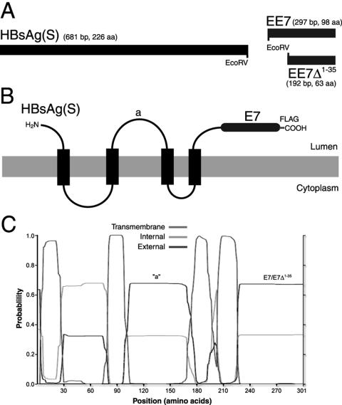
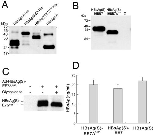
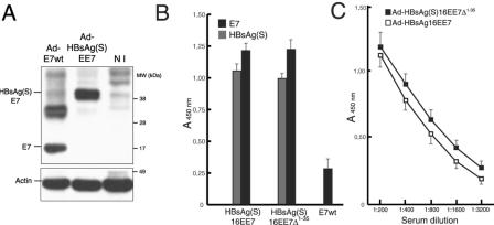
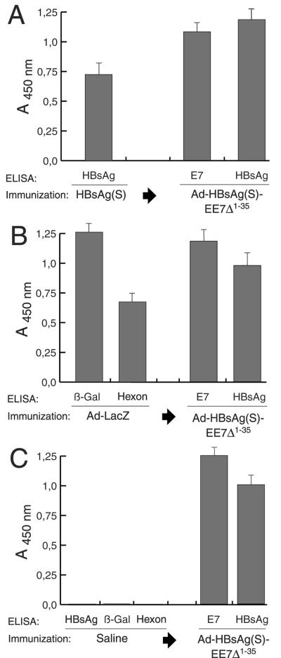
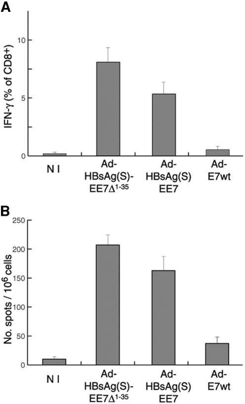
Induction of effective immune responses may help prevent cancer progression. Tumor-specific antigens, such as those of human papillomaviruses involved in cervical cancer, are targets with limited intrinsic immunogenicity. Here we show that immunization with low doses (10(6) infectious units/dose) of a recombinant human adenovirus type 5 encoding a fusion of the E7 oncoprotein of human papillomavirus type 16 to the carboxyl terminus of the surface antigen of hepatitis B virus (HBsAg) induces remarkable E7-specific humoral and cellular immune responses. The HBsAg/E7 fusion protein assembled efficiently into virus-like particles, which stimulated antibody responses against both carrier and foreign antigens, and evoked antigen-specific kill of an indicator cell population in vivo. Antibody and T-cell responses were significantly higher than those induced by a control adenovirus vector expressing wild-type E7. Such responses were not affected by preexisting immunity against either HBsAg or adenovirus. These data demonstrate that the presence of E7 on HBsAg particles does not interfere with particle secretion, as it occurs with bigger proteins fused to the C terminus of HBsAg, and results in enhancement of CD8(+)-mediated T-cell responses to E7. Thus, fusion to HBsAg is a convenient strategy for developing cervical cancer therapeutic vaccines, since it enhances the immunogenicity of E7 while turning it into an innocuous secreted fusion protein.


Figures

FIG. 1.
FIG. 1.
Schematic representation of HBsAg(S)16EE7 fusion genes and proteins and their expression in mammalian cells. (A) HBsAg(S)/E7 fusion genes used in this study. The stop codon of HBsAg(S) was replaced by an EcoRV site, which allows for in-frame 3′ end fusion of coding sequences. Constructs were derived by fusion of HBsAg(S) to either a complete EE7 gene or a truncated mutant devoid of the first 35 codons (EE7Δ1-35). A FLAG tag (not represented) was added at the 3′ end of the EE7 sequences. (B) Topology of the HBsAg(S)-HPV-16E7 fusion proteins in the ER membrane. The luminal side of the protein is that exposed on the surface of the extruded particles. (C) Transmembrane domain prediction of the HBsAg-E7 fusion proteins according to the algorithm developed by Sonnhammer et al. (37). The positions of the “a” determinant and the E7 domains are indicated.
FIG. 2.
FIG. 2.
Expression and secretion of HBsAg/E7 fusion proteins. (A) Expression of HBsAg/E7 fusion proteins in yeast. Western blotting using anti-HBsAg antibodies was carried out to detect His-tagged wild-type and chimeric HBsAg(S) proteins purified from yeast on Ni-nitrilotriacetic acid agarose. A wild-type HBsAg(S) with no His tag (Engerix B) was included. Note the higher molecular mass of HBsAg(S)-His compared to the nontagged protein. (B) Western blot analysis of HEK 293T cells transfected with pIRES-neo2 plasmids encoding FLAG-tagged HBsAg(S)16E7 or HBsAg(S)16E7Δ1-35, as indicated. Cellular extracts prepared 48 h after transfection were separated in a 15% acrylamide gel and hybridized with anti-FLAG M2 monoclonal antibodies. Note the thickness of the bands due to the presence of glycosylated and nonglycosylated forms running close to each other. The apparent molecular masses are 40 and 33 kDa, respectively. C, control cells transfected with pIRES-neo2 empty vector. (C) The HBsAg(S)/E7 fusion protein is glycosylated. Western blot analysis of lysates from HEK 293T cells infected with Ad-HBsAg(S)EE7Δ1-35 before and after treatment with Endo H and detected with anti-FLAG antibodies is shown. (D) Detection of HBsAg particles in supernatants of HeLa cells transfected with plasmids encoding either HBsAg(S)16E7, HBsAg(S)16E7Δ1-35 or wild-type HBsAg(S). Transfection efficiencies were normalized by cotransfection with a plasmid expressing green fluorescent protein. At 48 h after transfection, cell supernatants were subjected to ELISA (Monolisa). A standard curve was obtained using known amounts of a commercial HBsAg(S) particle preparation (Engerix B). Data are the means ± standard deviations of three independent transfections.
FIG. 3.
FIG. 3.
Transmission electron microscopy analysis of Vero cells transfected to express HBsAg(S)16E7Δ1-35. (a) Cells transfected with pIRESneo2-HBsAg(S)16EE7Δ1-35. This section corresponds to a low plane where the cell contains a number of large vacuolated structures surrounding the nucleus. Boxed area is magnified in panel b showing particles being extruded (arrowheads) into the lumen of a larger structure that contains a great number of particles (arrows). (c) A vesicle underneath the plasma membrane releasing its particle content to the extracellular medium. Li, lipid droplets; Mi, mitochondria; N, nucleus; Nu, nucleolus; V, large vacuoles containing 22-nm VLPs. Bar, 2 μm (a), 100 nm (b), and 225 nm (c).
FIG. 4.
FIG. 4.
Intracellular localization of the HBsAg(S)16E7 fusion protein analyzed by confocal microscopy. A representative Vero cell expressing HBsAg(S)16EE7 stained with an anti-FLAG antibody (green). Cells were counterstained with propidium iodide (red). Each photograph represents a single confocal section within a series in the xy and xz planes. (A) Three consecutive sections in the xy plane through the basal (xy1), medial (xy2), and apical (xy3) levels. The HBsAg(S)16E7 protein accumulates in large perinuclear structures derived from the ER and Golgi. In addition, a punctuate pattern of smaller vesicles containing fusion protein is seen scattered throughout the cytoplasm. These vesicles appear to migrate to the periphery, where they are exocytosed. Arrowheads point to HBsAg(S)16E7 protein into vesicular structures in neighboring untransfected cells. These are particle aggregates likely coming from the cell in the center, which was the only one transfected in this field. (B) Three cross-sections of the cells in the plane xz (perpendicular to the plane of adherence) at the levels indicated in xy2, showing large stained structures that are in tight contact with the nucleus and small vesicles that migrate distally to the periphery of the cell. The arrowhead in xz3 points to HBsAg(S)16E7 fusion protein captured by an untransfected cell. Bar, 20 μm.
FIG. 5.
FIG. 5.
Antibody reactivity to E7 and HBsAg(S) in vaccinated mice determined by ELISA. (A) Expression of E7 and HBsAg(S)E7 in HEK 293 cells infected with either Ad-E7wt (MOI of 100) or Ad-HBsAg(S)EE7 (MOI of 10). At 48 h after infection, the cells were lysed with SDS loading buffer and equal amounts were loaded on a 4 to 20% gradient gel. After blotting, the E7 and HBsAg(S)E7 proteins were detected with anti-E7 and anti-actin antibodies. The upper band in the lane Ad-E7wt represents likely a dimer. NI, noninfected control cells. (B) Groups of BALB/c mice were inoculated at 2-week intervals with 106 IFU of either Ad-HBsAg(S)16EE7, Ad-HBsAg(S)16EE7Δ1-35 or Ad-E7wt, as indicated. Results correspond to serum samples taken 2 weeks after the third inoculation. The sera were diluted 1:200 and assayed on plates coated with either recombinant E7 or HBsAg protein as described in Materials and Methods. Data are the means ± standard deviations of 10 serum samples. (C) Extinction curves of anti-E7 antibody titers of sera derived from the mice immunized with Ad-HBsAg(S)16EE7 or Ad-HBsAg(S)16EE7Δ1-35 vectors in panel A showing slightly higher titer values for the latter and extinction values for both beyond a dilution of 1:3,200. The ELISA plates were coated with the indicated proteins as described in Materials and Methods. The results show the mean OD and the standard deviation.
FIG. 6.
FIG. 6.
Antibody reactivity to E7 and HBsAg(S) in mice preimmunized with HBsAg or Ad-LacZ determined by ELISA. (A) BALB/c mice (n = 10) were first immunized i.m. with HBsAg(S) protein. Serum reactivity to HBsAg(S) was tested after 2 weeks after the third boost (left column). Subsequently, the mice were inoculated three times at 2-week intervals with Ad-HBsAg(S)16EE7Δ1-35. Serum samples were taken 2 weeks after the last inoculation and antibody titers against E7 and HBsAg were tested. (B) Vaccination of C57BL/6 mice (n = 10) preimmunized with Ad-LacZ. Mice were inoculated with Ad-LacZ at days 0, 14, and 28, beginning 2 weeks after the last boost antibody responses to the β-galactosidase and hexon proteins were tested. Subsequent vaccination with Ad-HBsAg(S)16EE7Δ1-35 induced additional antibody response against E7 and boosted the anti-HBsAg(S) (third and fourth columns from the left, respectively) (compare with panels A and C). (C) A control group of 10 C57BL/6 mice were injected with saline three times at 2-week intervals. Two weeks after the last injection the mice were tested for HBsAg, β-galactosidase, and Ad hexon antibodies. Then the mice were inoculated with Ad-HBsAg(S)16EE7Δ1-35 and tested as above for E7 and HBsAg antibodies. ELISA plates were coated with the indicated proteins as described in Materials and Methods. Data are the mean of 10 serum samples; the error bars represent one standard deviation from the mean values. Sera were diluted 1:200, and the cutoff value was 0.045 throughout.
FIG. 7.
FIG. 7.
Cytotoxic responses induced in C57BL/6 mice vaccinated with Ad vectors. (A) Intracellular flow cytometry analysis of E7-specific CD8+ T cells. Mice (n = 10 per group) were immunized with Ad vectors encoding HBsAg/E7 fusion proteins or Ad-E7wt. Splenocytes were restimulated for 7 days with E7(49-57) peptide. Intracellular cytokine staining was performed after a 6-h block with brefeldin A in the presence of E7(49-57) and anti-CD28 and anti-CD49d costimulator molecules. Nonstimulated controls were treated similarly but in the absence of E7(49-57). CD8+ lymphocytes were gated and analyzed by fluorescence-activated cell sorting for IFN-γ production. Percentages of IFN-γ-positive CD8+ cells are given. NI, nonimmunized control group. Nonstimulated cells gave values similar to those of the nonimmunized group. (B) T-cell response determined by IFN-γ ELISPOT of splenocytes from mice immunized with the indicated Ad vectors after overnight restimulation in vitro with E7(49-57) peptide. NI, nonimmunized control. Reactivity values from nonstimulated samples have been subtracted. Data are the means ± standard deviations of three independent assays.
FIG. 8.
FIG. 8.
Cytotoxic responses induced by vaccination with Ad encoding HBsAg/E7 fusion proteins determined by antigen-specific kill of indicator cell populations. C57BL/6 mice (n = 10 per group) were immunized at 2-week intervals with Ad-HBsAg(S)16EE7Δ49-57, Ad-16E7wt, or recombinant HBsAg(S)16E7Δ49-57 VLPs. Six days after the fourth boost immunization, splenocytes from naïve mice were loaded with either E7(49-57) or HBsAg(S)(208-215) peptides or left unloaded and subsequently labeled with CFSE to low, high, and intermediate concentrations, respectively. Equal amounts of labeled cells were mixed, and 15 × 106 cells of the mixture was injected intravenously into control naïve and vaccinated mice. Lymphocytes from spleens and regional lymph nodes were taken 20 h after injection and analyzed by flow cytometry. Represented are the mean percentages ± standard deviations of specific CTL activities in the different groups of mice calculated as described in Materials and Methods.

References

    1. Aichele, P., K. Brduscha-Riem, S. Oehen, B. Odermatt, R. M. Zinkernagel, H. Hengartner, and H. Pircher. 1997. Peptide antigen treatment of naive and virus-immune mice: antigen-specific tolerance versus immunopathology. Immunity 6:519-529. - PubMed
    1. Bontkes, H. J., T. D. de Gruijl, A. J. van den Muysenberg, R. H. Verheijen, M. J. Stukart, C. J. Meijer, R. J. Scheper, S. N. Stacey, M. F. Duggan Keen, P. L. Stern, S. Man, L. K. Borysiewicz, and J. M. Walboomers. 2000. Human papillomavirus type 16 E6/E7-specific cytotoxic T lymphocytes in women with cervical neoplasia. Int. J. Cancer. 88:92-98. - PubMed
    1. Bruss, V., and D. Ganem. 1991. Mutational analysis of hepatitis B surface antigen particle assembly and secretion. J. Virol. 65:3813-3820. - PMC - PubMed
    1. Cid-Arregui, A., V. Juarez, and H. zur Hausen. 2003. A synthetic E7 gene of human papillomavirus type 16 that yields enhanced expression of the protein in mammalian cells and is useful for DNA immunization studies. J. Virol. 77:4928-4937. - PMC - PubMed
    1. Davidson, E. J., R. L. Faulkner, P. Sehr, M. Pawlita, L. J. Smyth, D. J. Burt, A. E. Tomlinson, J. Hickling, H. C. Kitchener, and P. L. Stern. 2004. Effect of TA-CIN (HPV 16 L2E6E7) booster immunisation in vulval intraepithelial neoplasia patients previously vaccinated with TA-HPV (vaccinia virus encoding HPV 16/18 E6E7). Vaccine 22:2722-2729. - PubMed

Publication types

MeSH terms