. 2005 Oct;79(20):12828-39.
doi: 10.1128/JVI.79.20.12828-12839.2005.

Inhibition of interferon-stimulated JAK-STAT signaling by a tick-borne flavivirus and identification of NS5 as an interferon antagonist

Affiliations

Inhibition of interferon-stimulated JAK-STAT signaling by a tick-borne flavivirus and identification of NS5 as an interferon antagonist

Sonja M Best et al. J Virol. 2005 Oct.

Abstract

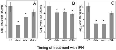
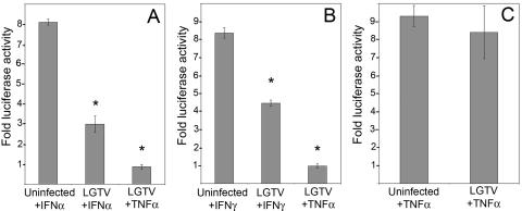
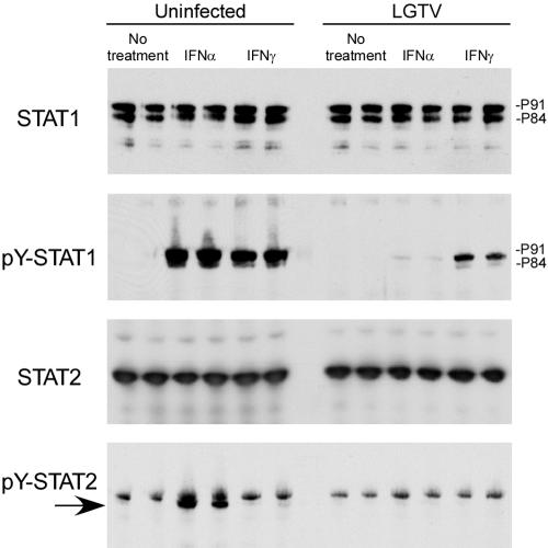
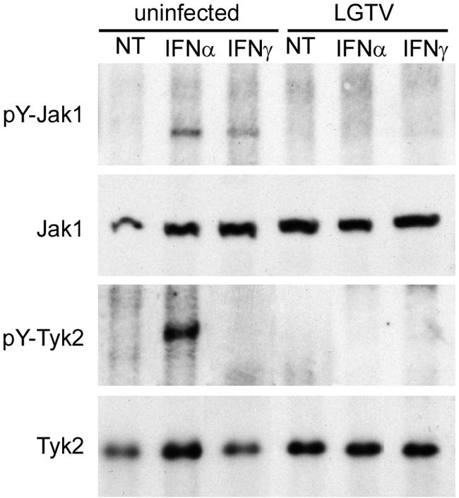
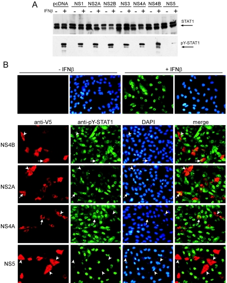
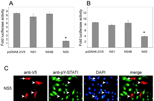
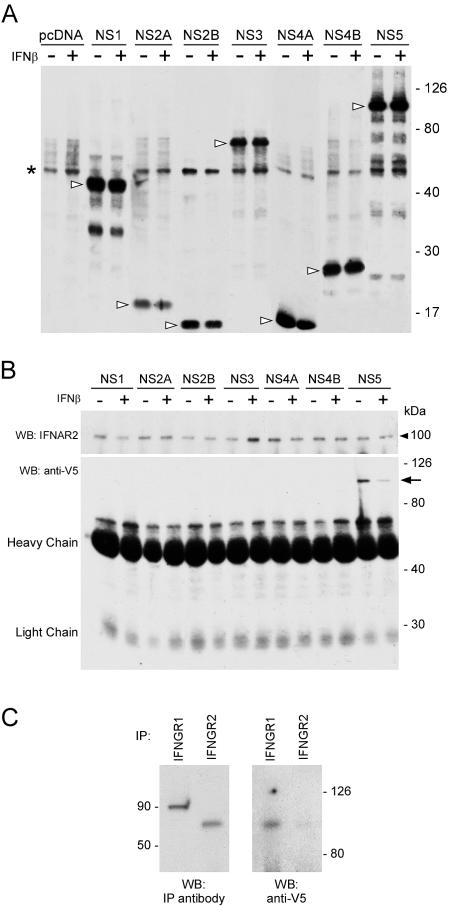
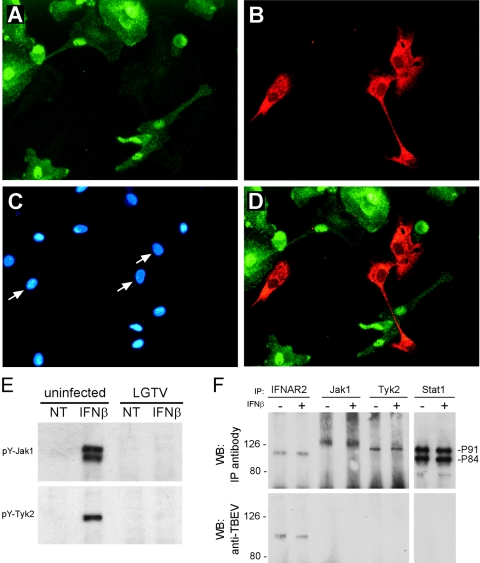
The tick-borne encephalitis (TBE) complex of viruses, genus Flavivirus, can cause severe encephalitis, meningitis, and/or hemorrhagic fevers. Effective interferon (IFN) responses are critical to recovery from infection with flaviviruses, and the mosquito-borne flaviviruses can inhibit this response. However, little is known about interactions between IFN signaling and TBE viruses. Langat virus (LGTV), a member of the TBE complex of viruses, was found to be highly sensitive to the antiviral effects of IFN. However, LGTV infection inhibited IFN-induced expression of a reporter gene driven by either IFN-alpha/beta- or IFN-gamma-responsive promoters. This indicated that LGTV can inhibit the IFN-mediated JAK-STAT (Janus kinase-signal transducer and activator of transcription) pathway of signal transduction. The mechanism of inhibition was due to blocks in the phosphorylation of both Janus kinases, Jak1 and Tyk2, during IFN-alpha signaling and at least a failure of Jak1 phosphorylation following IFN-gamma stimulation. To determine the viral protein(s) responsible, we individually expressed all nonstructural (NS) proteins and examined their ability to inhibit signal transduction. Expression of NS5 alone inhibited STAT1 phosphorylation in response to IFN, thus identifying NS5 as a potential IFN antagonist. Examination of interactions between NS5 and cellular proteins revealed that NS5 associated with IFN-alpha/beta and -gamma receptor complexes. Importantly, inhibition of JAK-STAT signaling and NS5-IFN receptor interactions were demonstrated in LGTV-infected human monocyte-derived dendritic cells, important target cells for early virus replication. Because NS5 may interfere with both innate and acquired immune responses to virus infection, this protein may have a significant role in viral pathogenesis.


Figures

FIG. 1.
FIG. 1.
Effect of IFN treatment on LGTV replication in MNB cells. MNB cells were infected with LGTV virus (strain TP21; MOI of 1) and incubated for 72 h, at which time the virus titer in the supernatant was determined. Cells were either not treated with IFN (NT), pretreated with IFN for 24 h (−24 hr), or treated with IFN 4 h (+4hr) or 24 h (+24hr) after infection. Cells were treated with (A) IFN-α (1,000 U/ml), (B) IFN-γ (10 ng/ml), or (C) IFN-α plus IFN-γ. The error bars represent standard errors of the mean (SEMs) for three experiments. *, P < 0.05.
FIG. 2.
FIG. 2.
Effect of LGTV (strain TP21) infection on luciferase reporter gene expression driven by ISRE, GAS, and NFκB promoters. Vero cells were transfected with (A) pISRE-luc, (B) pGAS-luc, or (C) pNFκB-luc reporter plasmids, infected with LGTV (MOI of 10) for 24 h, and treated with IFN-α (1,000 U/ml), IFN-γ (10 ng/ml), or TNF-α (100 μg/ml) for 6 to 7 h. Results are expressed as n-fold increases in luciferase activity over that of identical cultures not treated with cytokines (means ± SEMs for four experiments; *, P < 0.05).
FIG. 3.
FIG. 3.
Inhibition of tyrosine phosphorylation of STAT1 and STAT2 in response to IFN-α and IFN-γ by LGTV (strain TP21). Uninfected Vero cells or cells infected with LGTV for 48 h were treated with IFN-α or IFN-γ for 15 min or left untreated. The cell lysates were examined by Western blotting (shown in duplicate) with antibodies to STAT1, Tyr701-phosphorylated STAT1 (pY-STAT1), STAT2, or Tyr689-phosphorylated STAT2 (pY-STAT2; arrow), as indicated to the left of each panel.
FIG. 4.
FIG. 4.
Inhibition of JAK-STAT signaling in response to IFN is associated with inhibition of Jak1 and Tyk2 phosphorylation. Vero cells were infected with LGTV strain TP21 for 48 h and treated with IFN-α or IFN-γ for 15 min. The cell lysates were examined by immunoprecipitation using antibodies to Tyr1022/Tyr1023-phosphorylated Jak1 (pY-Jak1) or Tyr1054/1055-phosphorylated Tyk2 (pY-Tyk2). The immunoprecipitates were analyzed by Western blotting using antibodies that recognize Jak1 or Tyk2. Whole-cell lysates were also examined by Western blotting to determine steady-state levels of Jak1 and Tyk2.
FIG. 5.
FIG. 5.
Identification of LGTV nonstructural proteins that function as IFN antagonists. (A) Vero cells expressing LGTV NS proteins fused to V5 epitope tags were treated with blasticidin to obtain a cell population, the majority of which expressed the individual NS protein indicated, or the backbone plasmid, pcDNA6.2/V5. Cells were then treated with IFN-β and examined for STAT1 phosphorylation by Western blot analysis. Steady-state STAT1 levels (upper panel) and tyrosine-phosphorylated STAT1 (pY-STAT1) (lower panel) are shown in cell populations expressing LGTV NS proteins and either left untreated (−) or treated (+) with IFN-β. (B) Vero cells were treated with IFN-β, fixed in methanol, and stained for anti-V5 (red) and anti-pY-STAT1 (green). Nuclei were counterstained with DAPI (blue). The upper panel demonstrates the localization of tyrosine-phosphorylated STAT1 in the nuclei of untransfected cells that were left untreated or treated with IFN-β. The lower panels represent cells that express NS4B, NS2A, NS4A, and NS5. Examples of V5-positive and either pY-STAT1-positive (arrows) or pY-STAT1-negative (arrowheads) cells are indicated.
FIG. 6.
FIG. 6.
Effect of LGTV NS proteins on luciferase reporter gene expression. Vero cells transfected with (A) pISRE-luc or (B) pGAS-luc were also transfected with the backbone plasmid, pcDNA6.2/V5, or with the NS1, NS4B, or NS5 expression plasmid and treated with IFN-α or IFN-γ. Results are expressed as n-fold increases in luciferase activity over that of identical cultures not treated with cytokines (means ± SEMs for four experiments; *, P < 0.05). (C) NS5-expressing Vero cells were treated with IFN-γ, fixed, and stained for anti-V5 (red) and anti-pY-STAT1 (green). Nuclei were counterstained with DAPI (blue). Examples of V5-positive and either pY-STAT1-positive (arrow) or pY-STAT1-negative (arrowheads) cells are indicated.
FIG. 7.
FIG. 7.
Identification of LGTV NS proteins that associate with the IFN-α/β or IFN-γ receptor complexes in unstimulated (−) and IFN-β-stimulated (+) cells. (A) Western blot probed with anti-V5 antibody demonstrating the expression level of each NS-V5 fusion protein (arrowheads). The asterisk denotes a nonspecific anti-V5-reacting band present in all cell lysates. (B) Cell lysates from Vero cells expressing LGTV NS fusion proteins were immunoprecipitated with an anti-IFNAR2 monoclonal antibody (upper panel). Anti-V5-reacting, coprecipitating protein was detected by Western blot analysis (lower panel). Only the NS5 fusion protein coprecipitated with IFNAR2 (arrow). (C) Cell lysates from Vero cells expressing NS5 fusion protein were immunoprecipitated with anti-IFNGR1 or anti-IFNGR2 polyclonal antibodies. Anti-V5-reacting, coprecipitating protein was detected by Western blot analysis. The approximate molecular mass (kDa) is indicated. WB, Western blot; IP, immunoprecipitation.
FIG. 8.
FIG. 8.
JAK-STAT signaling is inhibited in LGTV-infected human monocyte-derived dendritic cells. Human monocyte-derived dendritic cells were infected with LGTV TP21 (MOI of 1) for 24 h prior to the addition of IFN-α for 15 min. Cells were fixed in methanol and stained for (A) pY-STAT1 and (B) LGTV E protein and (C) counterstained with DAPI to visualize nuclei. Arrows in panel C indicate the nuclei of cells infected with LGTV. (D) Merged image of panels A and B showing no pY-STAT1 in LGTV-infected dendritic cells. (E) Monocyte-derived DC were infected with LGTV TP21 for 48 h and treated with IFN-α for 15 min. The cell lysates were immunoprecipitated using antibodies to Tyr1022/Tyr1023-phosphorylated Jak1 (pY-Jak1) or Tyr1054/1055-phosphorylated Tyk2 (pY-Tyk2) and analyzed by Western blotting using the same antibodies. (F) Cell lysates from human DC infected for 48 h with LGTV, and either left untreated (−) or treated (+) with IFN-β, were immunoprecipitated with antibodies specific for IFNAR2, Jak1, Tyk2, or STAT1 (upper panels). Western blots examining coprecipitating LGTV proteins were probed with anti-TBEV antisera, revealing an association between NS5 and IFNAR2. WB, Western blot; IP, immunoprecipitation.

References

    1. Alexander, W. S., and D. J. Hilton. 2004. The role of suppressors of cytokine signaling (SOCS) proteins in regulation of the immune response. Annu. Rev. Immunol. 22:503-529. - PubMed
    1. Biron, C. A. 2001. Interferons alpha and beta as immune regulators-a new look. Immunity 14:661-664. - PubMed
    1. Bradford, M. M. 1976. A rapid and sensitive method for the quantitation of microgram quantities of protein utilizing the principle of protein-dye binding. Anal. Biochem. 72:248-254. - PubMed
    1. Brierley, M. M., and E. N. Fish. 2002. Review: IFN-alpha/beta receptor interactions to biologic outcomes: understanding the circuitry. J. Interferon Cytokine Res. 22:835-845. - PubMed
    1. Campbell, M. S., and A. G. Pletnev. 2000. Infectious cDNA clones of Langat tick-borne flavivirus that differ from their parent in peripheral neurovirulence. Virology 269:225-237. - PubMed

MeSH terms

LinkOut - more resources