. 2005 Oct;79(20):12905-13.
doi: 10.1128/JVI.79.20.12905-12913.2005.

Structural genomics of the severe acute respiratory syndrome coronavirus: nuclear magnetic resonance structure of the protein nsP7

Affiliations

Structural genomics of the severe acute respiratory syndrome coronavirus: nuclear magnetic resonance structure of the protein nsP7

Wolfgang Peti et al. J Virol. 2005 Oct.

Abstract

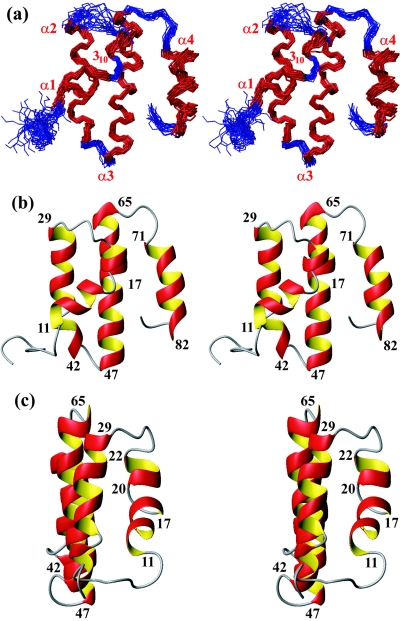
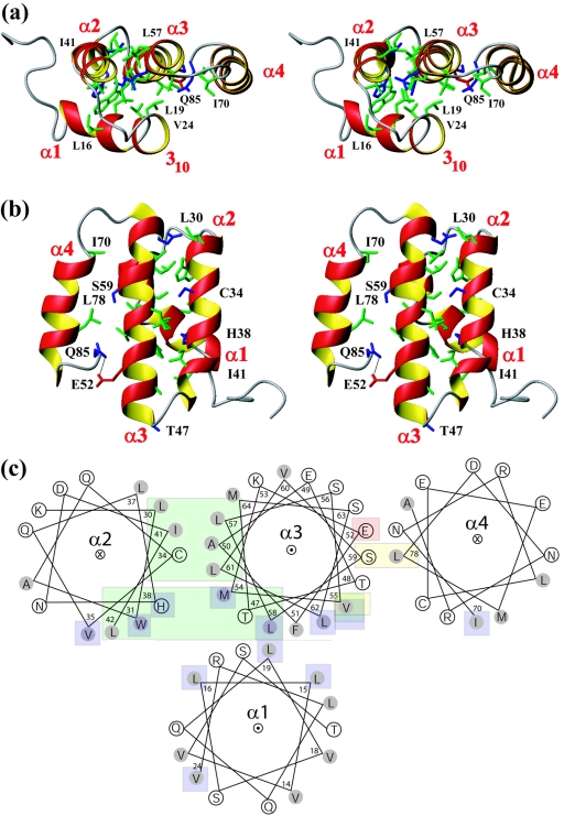
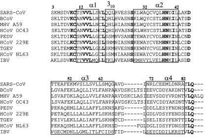
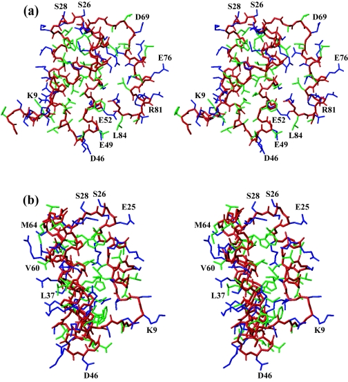
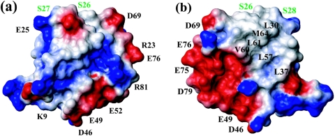
Here, we report the three-dimensional structure of severe acute respiratory syndrome coronavirus (SARS-CoV) nsP7, a component of the SARS-CoV replicase polyprotein. The coronavirus replicase carries out regulatory tasks involved in the maintenance, transcription, and replication of the coronavirus genome. nsP7 was found to assume a compact architecture in solution, which is comprised primarily of helical secondary structures. Three helices (alpha2 to alpha4) form a flat up-down-up antiparallel alpha-helix sheet. The N-terminal segment of residues 1 to 22, containing two turns of alpha-helix and one turn of 3(10)-helix, is packed across the surface of alpha2 and alpha3 in the helix sheet, with the alpha-helical region oriented at a 60 degrees angle relative to alpha2 and alpha3. The surface charge distribution is pronouncedly asymmetrical, with the flat surface of the helical sheet showing a large negatively charged region adjacent to a large hydrophobic patch and the opposite side containing a positively charged groove that extends along the helix alpha1. Each of these three areas is thus implicated as a potential site for protein-protein interactions.


Figures

FIG. 1.
FIG. 1.
Wall-eye stereo views of the solution structure of nsP7. (a) Bundle of the best 20 DYANA conformers of nsP7 after energy minimization. Only the polypeptide backbone is displayed. The 20 conformers have been superimposed for minimal RMSD of the backbone heavy atoms of residues 6 to 83. The four major helices, α1 to α4, and the 310-helix are colored red and labeled at their N termini. (b) Ribbon presentation of the closest conformer of nsP7 to the mean coordinates of the bundle shown in panel a, shown at the same viewing angle as for panel a. The sequence positions at both ends of the four α-helices are identified. (c) Same as panel b after rotation about a vertical axis, such that one looks at the edge of the up-down-up antiparallel three-α-helix sheet. The sequence positions at both ends of the helices α1, 310, α2, and α3 are identified.
FIG. 2.
FIG. 2.
(a) Top view of nsP7, generated from Fig. 1b by a 90° rotation about a horizontal axis in the projection plane. The helices are shown in a ribbon representation and are labeled at their N termini. Side chains involved in interhelix interactions (see the text) are shown as stick representations using the following color code: green, Ala, Leu, Val, Ile, Tyr, Trp, and Phe; blue, Ser, Thr, Cys, His, Asn, and Gln; red, Arg, Lys, Asp, and Glu. Some of the side chains discussed in the text are identified with the one-letter amino acid code and the residue number. (b) View of nsP7, generated from Fig. 1b by a 180° rotation about a vertical axis; the presentation is the same as for panel a. Some of the side chains discussed in the text are labeled, and a hydrogen bond between Glu 52 and Gln 85 is shown as a thin black line. Panels a and b show wall-eye stereo views. (c) Schematic top view of nsP7 (same as for panel a) with the helices represented as helical wheels. The helices α2 and α4 are directed from N to C into the page, and α3 runs out of the plane toward the viewer. α1 and the 310-helical turn following it are represented as one helical wheel running out of the page; this presentation does not show the tilt of α1 relative to the other three helices (Fig. 1). The side chains are represented as circles, with the hydrophobic side chains shaded. The side chains involved in α2-α3 interactions are shown on a green background, α3-α4 interactions are on a yellow background, and α1-α2, α3 interactions are on a blue background. Glu 52 is shown on a red background to indicate the hydrogen bonding interaction with the C-terminal residue, Gln 85. The helical wheel plot was prepared with the Web-based tool at http://kael.net.
FIG. 3.
FIG. 3.
Multiple alignment of coronavirus nsP7 sequences. Strictly conserved positions are indicated in boldface type. The positions of the helices α1 to α4 and 310 are indicated by boxes. Abbreviations and GenBank accession codes for the nsP7 sequences used are as follows: SARS-CoV, SARS coronavirus, strain Tor2, NP_828865; PEDV, porcine epidemic diarrhea virus, strain CV777, NP_839961; HCoV 229E, human coronavirus 229E, NP_835348; TGEV, transmissible gastroenteritis virus, strain Purdue, NP_840005; BCoV, bovine coronavirus ENT, NP_742134; MHV A59, murine hepatitis virus, strain A59, NP_740612; HCoV OC43, human coronavirus OC43, strain ATCC VR-759, NP_937947; HCoV NL63, human coronavirus NL63 strain Amsterdam I, YP_003766; IBV, avian infectious bronchitis virus, strain Beaudette, NP_740625. The residue numbering of nsP7 is according to the construct used in this study (see text), with the nsP7 sequence in positions 3 to 85. The sequence positions 3, 12, 22, 32, etc. are labeled (corresponding to residues 1, 10, 20, 30, etc. of the nsP7 sequence).
FIG. 4.
FIG. 4.
Wall-eye stereo views of all-heavy-atom presentations of the same conformer of nsP7 as that shown in Fig. 1b. (a) Viewing angle as in Fig. 1b. (b) Viewing angle as in Fig. 1c. Color code: green, hydrophobic side chains; blue, all other side chains; red, polypeptide backbone. Some of the side chains contributing to surface features discussed in the text are identified with the one-letter amino acid code and the residue number.
FIG. 5.
FIG. 5.
Surface views of nsP7 in a space-filling presentation. (a) Same orientation as in Fig. 1b. (b) View after a 180° rotation about a vertical axis, showing the surface formed by the flat three-helix sheet. Color code: gray, hydrophobic and polar residues; red, negatively charged; blue, positively charged. Some of the surface side chains discussed in the text are identified with the one-letter amino acid code and the residue number. The residues 26 to 28, which are discussed in the text as a putative functional site, are identified in green.

Similar articles

Cited by

References

    1. Alcami, A., and U. H. Koszinowski. 2000. Viral mechanisms of immune evasion. Immunol. Today 21:447-455. - PMC - PubMed
    1. Bost, A. G., R. H. Carnahan, X. T. Lu, and M. R. Denison. 2000. Four proteins processed from the replicase gene polyprotein of mouse hepatitis virus colocalize in the cell periphery and adjacent to sites of virion assembly. J. Virol. 74:3379-3387. - PMC - PubMed
    1. Bowie, A. G., J. Zhan, and W. L. Marshall. 2004. Viral appropriation of apoptotic and NF-κB signaling pathways. J. Cell. Biochem. 91:1099-1108. - PubMed
    1. Brockway, S. M., X. T. Lu, T. R. Peters, T. S. Dermody, and M. R. Denison. 2004. Intracellular localization and protein interactions of the gene 1 protein p28 during mouse hepatitis virus replication. J. Virol. 78:11551-11562. - PMC - PubMed
    1. Cornell, W. D., P. Cieplak, C. I. Bayly, I. R. Gould, K. M. Merz, Jr., D. M. Ferguson, D. C. Spellmeyer, T. Fox, J. W. Caldwell, and P. A. Kollman. 1995. A second generation force field for the simulation of proteins, nucleic acids, and organic molecules. J. Am. Chem. Soc. 117:5179-5197.

Publication types

MeSH terms

Associated data

LinkOut - more resources