. 2005 Oct;79(20):12914-20.
doi: 10.1128/JVI.79.20.12914-12920.2005.

Hepatitis B virus large and middle glycoproteins are degraded by a proteasome pathway in glucosidase-inhibited cells but not in cells with functional glucosidase enzyme

Affiliations

Hepatitis B virus large and middle glycoproteins are degraded by a proteasome pathway in glucosidase-inhibited cells but not in cells with functional glucosidase enzyme

Ender Simsek et al. J Virol. 2005 Oct.

Abstract

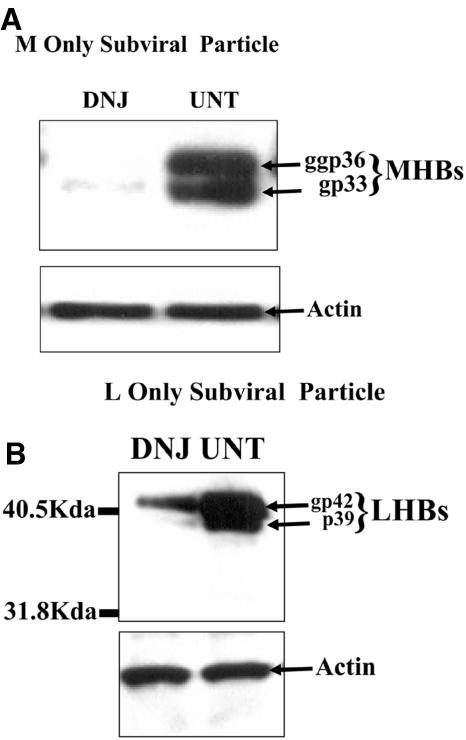
The secretion of hepatitis B virus (HBV) large (LHBs) and middle (MHBs) envelope polypeptides from tissue cultures requires proper protein folding and is prevented by inhibitors of the endoplasmic reticulum (ER) glucosidase. Using competitive inhibitors of the ER glucosidase, here it is shown that the amounts of glycosylated and unglycosylated forms of LHBs and MHBs proteins are all greatly reduced in tissue cultures producing HBV envelope glycoproteins. In contrast, the HBV small (SHBs) protein was not affected. The reduction in secretion of LHBs and MHBs proteins appears to be mediated by proteasomal degradation pathways, since it is prevented by either lactacystin or epoxomicin, two inhibitors of proteasomal degradation. Although there is no detectable proteasomal degradation of LHBs and MHBs in cells with functional glucosidase, the implications of the nearly quantitative sensitivity of glycosylated and unglycosylated forms of LHBs and MHBs proteins, with selective sparing of SHBs protein, in cells in which glucosidase is inhibited is surprising, and its implications are discussed.


Figures

FIG. 1.
FIG. 1.
Detection of HBV polypeptides LHBs and MHBs in HepG2 2.2.15 cell lysates as a function of glucosidase inhibition. (A) After 6 days of culture in the absence (UNT) or presence (DNJ) of the imino sugar glucosidase inhibitor DNJ, HBV polypeptides were detected in lysates following resolution through 12% SDS-PAGE using a polyclonal antibody that recognizes an epitope within pre-S2, which is in both LHBs and MHBs. Polypeptides corresponding to the molecular masses associated with LHBs and MHBs are indicated. The lower panel of panel A shows the same blot as that in the upper panel (washed) probed with a monoclonal antibody specific for actin. Panel B shows immunoblotting with the HBV-specific polyclonal antibody following resolution of an aliquot of lysate from the UNT samples before (−) and after (+) PNGase digestion. (C) The amount of LDH released in untreated cells is similar to that in drug-treated cells. This shows there is no toxicity in glucosidase-inhibited cells.
FIG. 2.
FIG. 2.
Secretion of HBV glycoproteins LHBs and MHBs from HepG2 2.2.15 cells as a function of glucosidase inhibition. After 6 days of culture in the absence (UNT) or presence (DNJ) of the glucosidase inhibitor deoxynojirimycin, the amounts of LHBs and MHBs were detected in the culture medium by Western blots, as in Fig. 1A. Polypeptides corresponding to the molecular massses of LHBs and MHBs are indicated. The presumed N-glycosylated species of LHBs (gpLHBs) and unglycosylated species (pLHBs) were resolved in this gel. (Lower panel) Immunoblot of the same blot shown (washed) in the upper panel, with monoclonal antibody specific for human albumin.
FIG. 3.
FIG. 3.
Secretion of AAT from HepG2 2.2.15 cells as a function of glucosidase inhibition. AAT secreted into the culture medium of 2.2.15 cells radiolabeled with [35S]Met, after 6 days of incubation in the presence (DNJ) and absence (UNT) of the glucosidase inhibitor DNJ, was detected by immunoprecipitation with monoclonal antibody. The autoradiograph of the immunoprecipitates resolved by SDS-PAGE is shown. Mature, 53-kDa ATT and presumed unprocessed species of AAT are indicated.
FIG. 4.
FIG. 4.
Accumulation and secretion of SHBs polypeptides from HepG2 2.2.15 cells as a function of glucosidase inhibition. (A) Lysates of HepG2 2.2.15 cells, maintained in the presence (DNJ) or absence (UNT) of glucosidase inhibitor DNJ for 6 days and labeled with [35S]methionine, as in Fig. 3, were immunoprecipitated and resolved by SDS-PAGE. The autoradiograph is shown and the bands corresponding to the SHBs and MHBs polypeptides are indicated. WB, Western blot of the same blot shown in the left panel, with polyclonal antibody specific for MHBs proteins. (B) The amount of SHBs in the culture medium (CM) or cell lysates (Cell) corresponding to the samples analyzed in panel A was determined with the Abbott Auszyme antigen capture assay, which uses a monoclonal antibody that recognizes the SHBs “a” epitope. Detection of antigen is reported as optical density (OD) values. UNT, cells left untreated; DNJ, cells incubated with glucosidase inhbitor DNJ.
FIG. 5.
FIG. 5.
Sensitivity of LHBs and MHBs to glucosidase inhibitors in the absence of other viral polypeptides. HepG2 cells were transfected with either plasmid pCMV-M, which specifies the HBV MHBs polypeptide in the absence of the other viral structural proteins (A), or pTRE-L, which specifies LHBs in the absence of the other viral structural proteins (B). Transfected cells were subcultured and further incubated for 6 days in the absence or presence of 4.45 mmol/liter imino sugar glucosidase inhibitor DNJ with HBV polypeptide (upper panels) or actin (lower panels) present in the cell lysate detected by Western blots using polyclonal antibodies, as described in the legends to the previous figures. The bands corresponding to the N-glycosylated species (gp) and unglycosylated species (p) are indicated, with these designations confirmed by PNGase digestion (not shown).
FIG. 6.
FIG. 6.
Evidence that functional proteasomes are necessary to mediate LHBs and MHBs sensitivity to glucosidase inhibitors. (A) Following incubation with the inhibitor DNJ (a glucosidase inhibitor) alone or in combination with either proteasome inhibitor LCT or EPO, LHBs and MHBs (upper panel) and actin (lower panel) were detected in cell lysates by Western blotting with specific polyclonal antibodies in cell lysates, as described previously for Fig. 1. Bands corresponding to the LHBs and MHBs polypeptides are indicated. The presence (+) or absence (−) of an inhibitor in a cell culture is indicated. (B) AAT was detected, by Western blotting, in lysates of HepG2 2.2.15 cells incubated in the absence (UNT) or presence of the glucosidase inhibitor DNJ. The lane under the label LCT UNT contains lysates from HepG2 2.2.15 cells incubated with LCT and no DNJ (UNT).

References

    1. Baumeister, W., J. Walz, F. Zuhl, and E. Seemuller. 1998. The proteasome: paradigm of a self-compartmentalizing protease. Cell 92:367-380. - PubMed
    1. Bergeron, J. J., M. B. Brenner, D. Y. Thomas, and D. B. Williams. 1994. Calnexin: a membrane-bound chaperone of the endoplasmic reticulum. Trends Biochem. Sci. 19:124-128. - PubMed
    1. Block, T. M., X. Lu, F. M. Platt, G. R. Foster, W. H. Gerlich, B. S. Blumberg, and R. A. Dwek. 1994. Hepatitis B virus secretion is inhibited by the imino sugar N-butyl-deoxynojirimycin. Proc. Natl. Acad. Sci. USA 91:2235-2239. - PMC - PubMed
    1. Block, T. M., X. Lu, A. S. Mehta, B. S. Blumberg, B. Tennant, M. Ebling, B. Korba, D. M. Lansky, G. S. Jacob, and R. A. Dwek. 1998. Treatment of chronic hepadnavirus infection in a woodchuck animal model with an inhibitor of protein folding and trafficking. Nat. Med. 4:610-614. - PubMed
    1. Block, T. M., and R. Jordan. 2002. Imino sugars as possible broad spectrum anti hepatitis virus agents: the alkovirs and glucovirs. Antiviral Chem. Chemother. 12:317-326. - PubMed

Publication types

MeSH terms

LinkOut - more resources