. 2005 Oct;79(20):13047-59.
doi: 10.1128/JVI.79.20.13047-13059.2005.

Herpes simplex virus 1 envelopment follows two diverse pathways

Affiliations

Herpes simplex virus 1 envelopment follows two diverse pathways

Helene Leuzinger et al. J Virol. 2005 Oct.

Abstract

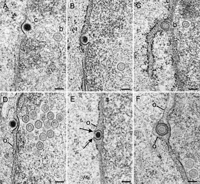
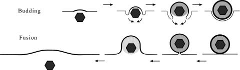
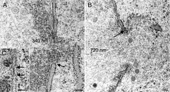
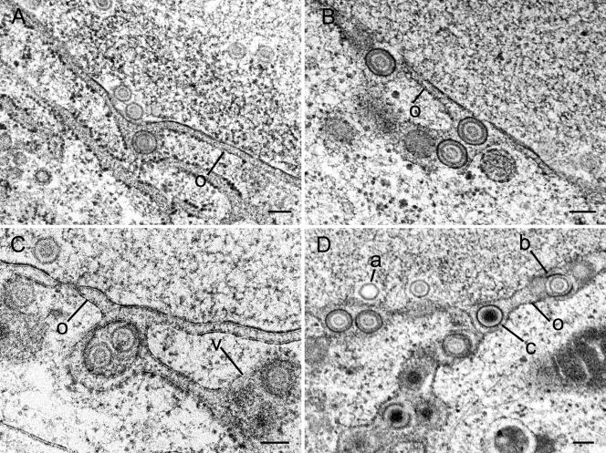
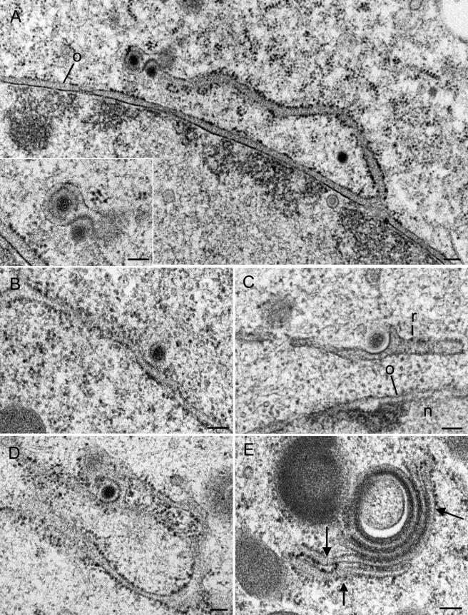
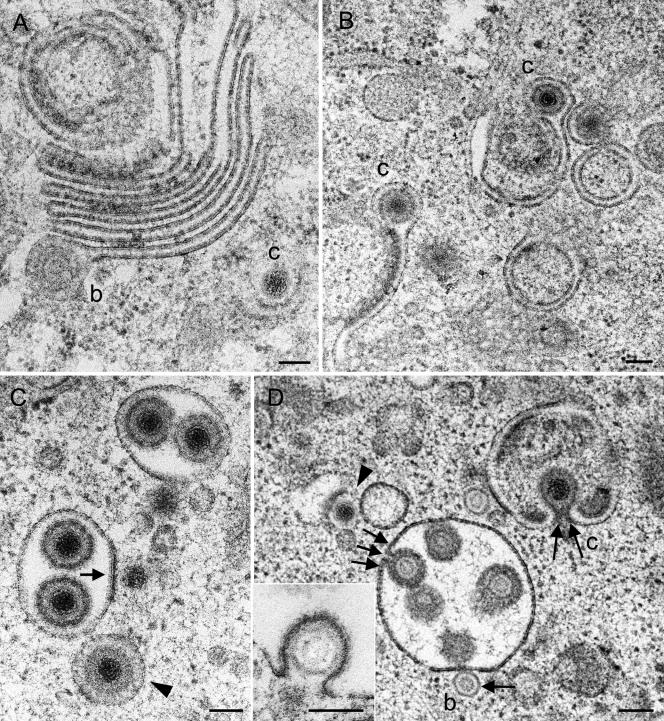
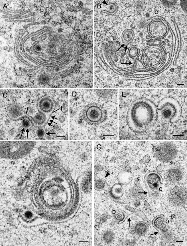
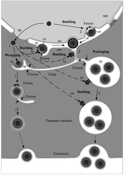
Herpesvirus envelopment is assumed to follow an uneconomical pathway including primary envelopment at the inner nuclear membrane, de-envelopment at the outer nuclear membrane, and reenvelopment at the trans-Golgi network. In contrast to the hypothesis of de-envelopment by fusion of the primary envelope with the outer nuclear membrane, virions were demonstrated to be transported from the perinuclear space to rough endoplasmic reticulum (RER) cisternae. Here we show by high-resolution microscopy that herpes simplex virus 1 envelopment follows two diverse pathways. First, nuclear envelopment includes budding of capsids at the inner nuclear membrane into the perinuclear space whereby tegument and a thick electron dense envelope are acquired. The substance responsible for the dense envelope is speculated to enable intraluminal transportation of virions via RER into Golgi cisternae. Within Golgi cisternae, virions are packaged into transport vacuoles containing one or several virions. Second, for cytoplasmic envelopment, capsids gain direct access from the nucleus to the cytoplasm via impaired nuclear pores. Cytoplasmic capsids could bud at the outer nuclear membrane, at membranes of RER, Golgi cisternae, and large vacuoles, and at banana-shaped membranous entities that were found to continue into Golgi membranes. Envelopes originating by budding at the outer nuclear membrane and RER membrane also acquire a dense substance. Budding at Golgi stacks, designated wrapping, results in single virions within small vacuoles that contain electron-dense substances between envelope and vacuolar membranes.


Figures

FIG. 1.
FIG. 1.
Budding capsids at inner and outer nuclear membranes in Vero and Vero 2-2 cells infected with HSV-1 at 10 h (B), 12 h (A, C, D, and E), and 15 h (F) postincubation. (A) C-capsid (c) at the beginning of budding at the inner nuclear membrane (i), which is thickened by an electron-dense substance at the site of budding. (B) Budding C-capsid with about three-fourths of the envelope being part of the inner nuclear membrane. Tegument is present only between the capsid and the thickened membrane. (C) Budding B-capsid (b), probably at the inner nuclear membrane, that is pushed into the perinuclear space at the site of an associated RER cisterna. (D) C-capsid at the beginning of budding at the outer nuclear membrane. Both the inner and outer nuclear membranes are thickened by an electron-dense substance. At the nuclear periphery is a small cluster of B-capsids. (E) C-capsid close to completion of budding at the outer nuclear membrane (o). The membrane (about four-fifths of the perimeter) around the capsid is thickened and turns in a sharp loop (arrows) into the normal outer nuclear membrane. (F) B-capsid within the perinuclear space containing tegument and a dense envelope, probably immediately after fission from the outer nuclear membrane, which is slightly indented. Bars, 100 nm.
FIG. 2.
FIG. 2.
Schematic drawing of budding and fusion. Budding induces thickening of the membrane that is pulled behind the capsid. The space between capsids and the envelope is progressively filled by tegument. The resulting virion is finally released by fission. Fusion starts by close apposition of the virion to the membrane followed by pore formation between the envelope and the membrane. Capsid and tegument are rapidly released, and the membrane is flattened because there are no forces to keep it bent. The thickened membrane would persist at the entire length equaling the viral surface.
FIG. 3.
FIG. 3.
Impairment of nuclear pores in Vero 2-2 cells and HeLa cells infected with HSV-1 after 12 h (A) and 14 h (B) of incubation. (A) Dilated nuclear pore with intact nuclear membrane (arrow) and nuclear substance protruding into the cytoplasm. (B) B-capsids escaping through an impaired nuclear pore with intact membrane at the border (arrow). (C) Intact nuclear pore of a mock-infected HeLa cell (17 h) with a distinctly visible nuclear pore complex and bordering membranes (arrows). Bars, 100 nm.
FIG. 4.
FIG. 4.
Confocal microscopy of HeLa cells after immunolabeling of pore complex protein Nup153 (Texas Red) and of HSV-1 immediate-early protein ICP4 (D) and subsequent DAPI staining. (A) Faint Texas Red signals are regularly distributed at the nuclear surface in cells incubated for 4 h with HSV-1. (B) Signal intensity is increased at the nuclear surface in cells incubated for 6 h. (C) Irregular accumulation of Texas Red signals at the nuclear periphery and in the cytoplasm at 8 h postinfection. (D) Merger of Texas Red, FITC, and DAPI staining demonstrating that dislocation of Nup153 from the nuclear periphery into the cytoplasm occurred only in cells expressing HSV-1 virus protein ICP4 (FITC). Bars, 5 μm.
FIG. 5.
FIG. 5.
Virions within perinuclear space and associated RER cisternae 12 h (B), 15 h (C), and 17 h (A and D) after infection of Vero and Vero 2-2 cells with HSV-1. (A to C) Single virions comprising one or two (C) B-capsids, tegument, and dense envelope within the perinuclear space (A) and associated RER cisternae (B and C) that continue into a vacuole-like structure (v) sectioned tangentionally in panel C. Note the dilation of the perinuclear space and RER at sites where virions are present. (D) Virions containing B- and C-capsids queue within the perinuclear space and associated RER cisternae. a, A-capsid; o, outer nuclear membrane. Bars, 100 nm.
FIG.6.
FIG.6.
Budding capsids in RER cisternae of Vero 2-2 cells at 12 h postinfection (A, C, and D) and of HeLa cells at 16 h postinfection (B and E) with HSV-1. (A) RER membrane continuous with the outer nuclear membrane (o) is associated with Golgi membranes surrounding a virion (inset). A cytoplasmic capsid is in an early phase of budding at the site where RER and Golgi membranes are associated. (B to D) Capsids in initial (B), early (C), and late (D) phases of budding into RER cisternae, for which the membrane situated close to the nucleus (n) has only a few ribosomes (r) in panel C. Note that all membrane capsid buds are thickened by a dense substance. (E) Golgi complex with narrow cisternae filled with a dense substance, probably tegument. Note the continua between Golgi and RER membranes (arrows). Bars, 100 nm.
FIG. 7.
FIG. 7.
Golgi complex and packaging-derived vacuoles or cross-sectioned Golgi cisternae in Vero 2-2 cells incubated for 12 h (A, B, and C) and 15 h (D) with HSV-1. (A) Large Golgi complex bearing a virion with a B-capsid (b) at the lateral end of a Golgi cisterna in a late stage of packaging. C-capsid (c) budding is shown at a banana-shaped membrane. (B) Two single Golgi cisternae with C-capsids late in packaging. (C) Two vacuoles derived from packaging or cross-sectioned Golgi cisternae, each containing two virions, and a vacuole (arrowhead) with a virion exactly in the center, with the space between the envelope and the vacuolar membrane containing a dense substance that might have resulted from wrapping or packaging. C-capsids are in close apposition to the packaging-derived vacuoles, one of which is in an initial phase of budding (arrow). (D) B-capsid in an initial phase (arrow) and C-capsid in a late phase (two arrows) of budding into vacuoles or cross-sectioned Golgi cisternae. One virion (three arrows) is shown either immediately prior to completion of budding or in an early stage of fusion. A C-capsid (arrowhead) in an early phase of budding at a banana-shaped membrane is shown. The inset shows a B-capsid close to completion of budding into a vacuole. Bars, 100 nm.
FIG.8.
FIG.8.
Budding capsids at Golgi membranes in Vero 2-2 cells at 12 h (A, C, F, and G) and in HeLa cells at 15 h (E) and 17 h (B and D) postinfection. (A) C-capsid at initial phase of wrapping at a slightly thickened membrane in a central region of a large Golgi complex. (B) Virions inside concentric vacuoles close to the completion of fission. The vacuolar membrane is still connected to the origin in the section plane (arrow) and probably below the section plane (two arrows). A C-capsid (c) approaches a large Golgi cisterna, and another capsid (arrowhead) is midway through being wrapped by a short Golgi cisterna. (C) C-capsids in initial (arrow), early (two arrows), and late (three arrows) phases of wrapping by a Golgi cisterna, from which two virions are dispatched by fission (f) as a result of packaging. (D) Virion with a dense envelope inside a concentric vacuole, possibly derived by wrapping because of the dense substance between the envelope and the vacuolar membrane. (E) C-capsid in an early phase of wrapping by a Golgi cisterna or Golgi-derived vacuole with a surface that is not sufficient to form the substance between the envelope and the surrounding vacuolar membrane, at least in this section plane. (F) C-capsid in an early phase of budding at a banana-shaped cisterna of a large Golgi complex. (G) C-capsid and two B-capsids budding at small membrane pieces that might be connected (arrows) to each other or to other membrane entities outside of this section plane. At the left is a tangentially cut virion within a Golgi cisterna (arrowhead). Bars, 100 nm.
FIG. 9.
FIG. 9.
Schematic drawing of the pathways of HSV-1 envelopment. (1) Cytoplasmic envelopment. Capsids leave the nucleus via impaired nuclear pores (np) and approach Golgi membranes from the cytoplasmic side, inducing budding. Since the entire cisterna is involved, a sphere-like structure comprising two membranes arises. The inner membrane becomes the viral envelope, and the outer one becomes the vacuolar membrane. Fusion of the envelope with the vacuolar membrane is considered likely to be prevented by “antifusion” proteins at high concentrations. Alternatively, capsids escaping the nucleus via impaired nuclear pores can also bud at the outer nuclear membrane (1a), RER membranes (1a), membranes of dilated Golgi cisternae (1b), and membranes of Golgi-derived vacuoles (1b) already containing virions. Virions originating by budding at the outer nuclear membrane and RER membranes need to be transported to Golgi cisternae for packaging. (2) Nuclear envelopment. Capsids bud through the inner nuclear membrane and are transported from the perinuclear space via RER cisternae into Golgi cisternae for packaging into transport vacuoles of various sizes containing one or more virions. Fusion of the viral envelope with cell membranes of the compartments the virion is transported through is considered likely to be prevented by proteins of an unknown nature, which are condensed (co) at the viral envelope. The “antifusion proteins” are speculated to be removed from the envelope in Golgi cisternae but are assumed to remain in the transport vacuoles (possibly together with additional proteins) at low concentrations to prevent the fusion of viral envelopes with the membranes of large transport vacuoles. im, inner nuclear membrane; om, outer nuclear membrane; npc, nuclear pore complex; sp, spikes; te, tegument.

Comment in

  • Egress of alphaherpesviruses.
    Mettenleiter TC, Minson T. Mettenleiter TC, et al. J Virol. 2006 Feb;80(3):1610-1; author reply 1611-2. doi: 10.1128/JVI.80.3.1610-1612.2006. J Virol. 2006. PMID: 16415038 Free PMC article. No abstract available.

Similar articles

Cited by

References

    1. Avitabile, E., G. Lombardi, T. Gianni, M. Capri, and G. Campadelli-Fiume. 2004. Coexpression of UL20p and gK inhibits cell-cell fusion mediated by herpes simplex virus glycoproteins gD, gH-gL, and wild-type gB or an endocytosis-defective gB mutant and downmodulates their cell surface expression. J. Virol. 78:8015-8025. - PMC - PubMed
    1. Baines, J. D., C. Cunningham, D. Nalwanga, and A. Davison. 1997. The U(L)15 gene of herpes simplex virus type 1 contains within its second exon a novel open reading frame that is translated in frame with the U(L)15 gene product. J. Virol. 71:2666-2673. - PMC - PubMed
    1. Borchers, K., and M. Oezel. 1993. Simian agent 8 (SA8): morphogenesis and ultrastructure. Zentbl. Bakteriol. 279:526-536. - PubMed
    1. Campadelli-Fiume, G., F. Farabegoli, S. Di Gaeta, and B. Roizman. 1991. Origin of unenveloped capsids in the cytoplasm of cells infected with herpes simplex virus 1. J. Virol. 65:1589-1595. - PMC - PubMed
    1. Card, J. P., L. Rinaman, R. B. Lynn, B. H. Lee, R. P. Meade, R. R. Miselis, and L. W. Enquist. 1993. Pseudorabies virus infection of the rat central nervous system: ultrastructural characterization of viral replication, transport, and pathogenesis. J. Neurosci. 13:2515-2539. - PMC - PubMed

Publication types

LinkOut - more resources