. 2005 Oct;79(20):13116-28.
doi: 10.1128/JVI.79.20.13116-13128.2005.

Hepatocyte-like cells transdifferentiated from a pancreatic origin can support replication of hepatitis B virus

Affiliations

Hepatocyte-like cells transdifferentiated from a pancreatic origin can support replication of hepatitis B virus

Robert Yung-Liang Wang et al. J Virol. 2005 Oct.

Abstract

Recently, a rat pancreatic cell line (AR42J-B13) was shown to transdifferentiate to hepatocyte-like cells upon induction with dexamethasone (Dex). The aim of this study is to determine whether transdifferentiated hepatocytes can indeed function like bona fide liver cells and support replication of hepatotropic hepatitis B virus (HBV). We stably transfected AR42J-B13 cells with HBV DNA and examined the expression of hepatocyte markers and viral activities in control and transdifferentiated cells. A full spectrum of HBV replicative intermediates, including covalently closed circular DNA (cccDNA) and Dane particles, were detected only after induction with Dex and oncostatin M. Strikingly, the small envelope protein and RNA of HBV were increased by 40- to 100-fold upon induction. When HBV RNAs were examined by primer extension analysis, novel core- and precore-specific transcripts were induced by Dex which initiated at nucleotide (nt) 1820 and nt 1789, respectively. Most surprisingly, another species of core-specific RNA, which initiates at nt 1825, is always present at almost equal intensity before and after Dex treatment, a result consistent with Northern blot analysis. The fact that HBV core protein is dramatically produced only after transdifferentiation suggests the possibility of both transcriptional and translational regulation of HBV core antigen in HBV-transfected AR42J-B13 cells. Upon withdrawal of Dex, HBV replication and gene expression decreased rapidly-less than 50% of the cccDNA remained detectable in 1.5 days. Our studies demonstrate that the transdifferentiated AR42J-B13 cells can function like bona fide hepatocytes. This system offers a new opportunity for basic research of virus-host interactions and pancreatic transdifferentiation.


Figures

FIG. 1.
FIG. 1.
Stable HBV-transfected clones B13-1 and B13-28 can be induced to secrete HBeAg (A) and HBsAg (B) by Dex and OSM. The ELISA procedure and data processing for HBsAg and HBeAg were according to the manufacturer's instructions (International Immunodiagnostics Co.). The values and standard deviations are from a total of four independent induction experiments. The results were calculated by means of a cutoff value determined with the following formula: negative control + 0.100 = cutoff (Co); S/Co = (sample signal optical density at 450 nm)/cutoff. Positive control, >3.000; negative control, 0.023. The accumulated levels of secreted HBsAg and HBeAg from B13-1 and B13-28 cells between day 5 and day 7 after the Dex plus OSM treatment are comparable to those accumulated for 48 h in the freshly changed medium from an HBV-producing Qs21 rat hepatoma cell line (49).
FIG. 2.
FIG. 2.
Immunofluorescence staining of HBV core antigen and liver-specific glutamine synthetase in transdifferentiated B13-1 and B13-28 cells. Cells were treated with Dex plus OSM for 7 days, followed by dual immunostaining with anti-HBc (green) and anti-glutamine synthetase (red). (A) B13-1 cells: a, anti-HBc; b, anti-glutamine synthetase; c, overlaid image of images from panels a and b; d, differential interference contrast (DIC); e, uninduced control stained with anti-HBc; f, DAPI staining for nuclei of the same field as in panel e. (B) B13-28 cells: g, anti-HBc; h, anti-glutamine synthetase; i, overlaid image of images from panels g and h; j, DIC; k, uninduced control, anti-glutamine synthetase; l, DAPI staining for nuclei of the same field as in panel k.
FIG. 3.
FIG. 3.
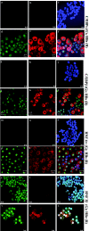
Immunofluorescence staining of HBV surface antigen and liver-specific transferrin in transdifferentiated B13-1 and B13-28 cells. (A) B13-1 cells were treated with Dex plus OSM for 5 (a to d) or 7 (e to h) days and then immunostained with anti-HBs (green) and anti-transferrin (red). (B) B13-28 cells were treated with Dex plus OSM for 5 (i to l) or 7 (m to p) days and then immunostained with anti-HBs (green) and anti-transferrin (red). The negative control results prior to induction (data not shown) were very similar to those in Fig. 2e and k.
FIG. 4.
FIG. 4.
Comparisons of the colocalization patterns of the liver-enriched transcription factors with HBsAg and HBcAg in transdifferentiated B13-28 cells. Cells were treated with or without Dex plus OSM for 7 days, followed by dual immunostaining. (a to c, g to i, m to o, and s to u) Before induction. a, anti-C/EBPα (green); b, anti-HBs (red); c, overlaid image of images a and b; g, anti-C/EBPβ (green); h, anti-HBs (red); i, overlaid image of images g and h; m, anti-HNF 4α (green); n, anti-HBs (red); o, overlaid image of images m and n; s, anti-HNF 3β (green); t, anti-HBc (red); u, overlaid image of images s and t. (d to f, j to l, p to r, and v to x) After induction. d, anti-C/EBPα (green); e, anti-HBs (red); f, overlaid image of images d and e; j, anti-C/EBPβ (green); k, anti-HBs (red); l, overlaid image of images j and k; p, anti-HNF 4α (green); q, anti-HBs (red); r, overlaid image of images p and q; v, anti-HNF 3β (green); w, anti-HBc (red); x, overlaid image of images v and w.
FIG. 5.
FIG. 5.
Western blot analysis of liver, pancreas, and HBV core protein expression in uninduced and induced B13-1 and B13-28 cells. Each lane on the sodium dodecyl sulfate-polyacrylamide gel electrophoresis was loaded with total proteins extracted from approximately one million cells. An enhanced chemiluminescence Western blotting detection kit was used as suggested by the vendor (Amersham Biosciences Co., United Kingdom).
FIG. 6.
FIG. 6.
(A) Production of HBV small envelope-specific mRNA (pre-S2/S) can be significantly stimulated by Dex plus OSM. Twenty-five micrograms of total RNA from each sample was analyzed by Northern blotting using a radiolabeled vector-free 3.1-kb HBV DNA probe. HBV RNA from Qs21 was included as a positive control. Major HBV-specific transcripts are indicated by arrows. 18S and 28S rRNA are shown below as an internal control. (B) Primer extension analysis revealed constitutive and Dex-inducible core- and precore-specific RNA species in B13-1 and B13-28 cells. Twenty-five micrograms of total RNAs, which were isolated from B13-1 and B13-28 cells with or without Dex plus OSM for 7days, was used as the template for primer extension analysis. The extended products corresponding to precore- and core-specific RNAs are indicated by arrows.
FIG. 7.
FIG. 7.
(A) Intracellular HBV DNA replication in B13-1 and B13-28 cells was significantly increased upon treatment with Dex plus OSM. Approximately 6 million cells from each 10-cm dish were harvested at different time points after treatment with Dex plus OSM for 3, 5, and 7 days. The letter U represents uninduced cells without Dex and OSM for 0 to 5 days. Qs21 is an HBV-producing cell line and was included here as a positive control. Each lane was loaded with HBV DNA extracted directly from cell lysates of each 10-cm dish. A 3.1-kb radiolabeled HBV DNA was used as a probe. (B) Southern blot analysis of ccc HBV DNA in transdifferentiated B13-1 and B13-28 cells. The cccDNA loaded in each lane was extracted from approximately 6 million cells from each 10-cm dish grown with Dex plus OSM for 7 to 9 days (see Materials and Methods). The strong banding intensity of the EcoRI-digested 3.2-kb fragment originates in part from the linearized cccDNA and in part from the trace amount of contaminating cellular DNA containing stably integrated HBV DNA concatemer. (C) Southern blot analysis of extracellular HBV DNA in the medium of transdifferentiated B13-28 cells. The signals in each lane correspond to approximately 20 ml of 48-h conditioned medium collected on days 5 and 7 postinduction. After centrifugation through a 20% sucrose cushion, the resuspended pellets of HBV particles were separated by isopycnic centrifugation through a cesium chloride gradient (20 to 50%). The fractions corresponding to Dane particles (fractions 10 to 16; density of 1.24 g/cm3) were pooled, and extracellular HBV DNA was extracted and subjected to Southern blot analysis.
FIG. 8.
FIG. 8.
Electron microscopic examination of secreted HBV viral and subviral particles in the medium of B13-28 cells induced with Dex plus OSM for 7 and 9 days. Particles are spherical (A) or filamentous (B) subviral particles and 42-nm Dane-like particles (C). On day 7 posttreatment with Dex-OSM, a total of 6 million B13-28 cells in one 10-cm dish were estimated to accumulate approximately 107 virion particles in 24 h (data not shown).
FIG. 9.
FIG. 9.
Continuous presence of Dex plus OSM is required for HBV replication and gene expression in B13-1 and B13-28 cells. Both B13-1 and B13-28 cells were treated with Dex plus OSM for 7 days. They were then cultured in the medium without Dex plus OSM for 3 or 7 days. The conditioned media were collected for ELISAs of HBsAg and HBcAg (A), and viral DNAs isolated from the total cells in each entire dish were harvested and loaded on each lane for Southern blot analyses (B and C). Rapid decline of HBV RC and SS DNA was detected after withdrawal (B). The time course of HBV cccDNA was measured on days 0, 1, 2, 3, and 5 after Dex withdrawal (C). (D) Western blot analysis for the proteins of HBcAg, α1-anti-trypsin, α-amylase, HBsAg, and α-tubulin.
FIG. 10.
FIG. 10.
While the expression of HNF 3β is independent of Dex plus OSM, the expression of C/EBPα, C/EBPβ, and HNF 4α declines rapidly upon withdrawal of Dex and OSM in transdifferentiated B13-28 cells. B13-28 cells were treated with Dex plus OSM for 7 days. They were then cultured in the medium without Dex plus OSM for 3 days, followed by dual immunostaining. A, anti-C/EBPα (green) and anti-HBs (red); B, anti-C/EBPβ (green) and anti-HBs (red); C, anti-HNF 4α (green) and anti-HBs (red); D, anti-HNF 3β (green) and anti-HBc (red). Bar, 10 μm.
FIG. 11.
FIG. 11.
Cartoon illustration of expression profiles of core-specific mRNA in B13-1 and B13-28 cells before and after Dex treatment. The first four patterns are predictions based on hypothetical mechanisms. P, pancreatic cells; H, hepatocytes.

Similar articles

Cited by

References

    1. Acs, G., M. S. Sells, R. H. Purcell, P. Price, R. Engle, M. Shapiro, and H. Popper. 1987. Hepatitis B virus produced by transfected Hep G2 cells causes hepatitis in chimpanzees. Proc. Natl. Acad. Sci. USA 84:4641-4644. - PMC - PubMed
    1. Addison, W. R., K. A. Walters, W. W. S. Wong, J. S. Wilson, D. Madej, L. D. Jewell, and D. L. J. Tyrrell. 2002. Half-life of the duck hepatitis B virus covalently closed circular DNA pool in vivo following inhibition of viral replication. J. Virol. 76:6356-6363. - PMC - PubMed
    1. Araki, K., J. Miyazaki, O. Hino, N. Tomita, O. Chisaka, K. Matsubara, and K. Yamamura. 1989. Expression and replication of hepatitis B virus genome in transgenic mice Proc. Natl. Acad. Sci. USA 86:207-211. - PMC - PubMed
    1. Babinet, C., H. Farza, D. Morello, M. Hadchouel, and C. Pourcel. 1985. Specific expression of hepatitis B surface antigen (HBsAg) in transgenic mice. Science 230:1160-1163. - PubMed
    1. Chang, C. M., K. S. Jeng, C. P. Hu, S. J. Lo, T. S. Su, L. P. Ting, C. K. Chou, S. H. Han, E. Pfaff, and J. Salfeld. 1987. Production of hepatitis B virus in vitro by transient expression of cloned HBV DNA in a hepatoma cell line. EMBO J. 6:675-680. - PMC - PubMed

Publication types