. 2005 Oct;49(10):4154-65.
doi: 10.1128/AAC.49.10.4154-4165.2005.

Resistance to antimicrobial peptides and stress response in Mycoplasma pulmonis

Affiliations

Resistance to antimicrobial peptides and stress response in Mycoplasma pulmonis

Lina Fassi Fehri et al. Antimicrob Agents Chemother. 2005 Oct.

Abstract

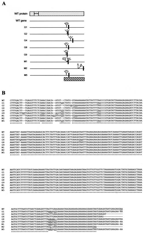
Antimicrobial peptides are widely distributed in nature, and in vertebrates, they play a key function in the innate immune defense system. It is generally agreed that these molecules may provide new antibiotics with therapeutic value. However, there are still many unsolved questions regarding the mechanisms underlying their antimicrobial activity as well as the mechanisms of resistance evolved by microorganisms against these molecules. The second point was addressed in this study. After determining the activity of 10 antimicrobial peptides against Mycoplasma pulmonis, a murine respiratory pathogen, the development of resistance was investigated. Following in vitro selection using subinhibitory concentrations of peptides, clones of this bacterium showing increased resistance to melittin or gramicidin D were obtained. For some of the clones, a cross-resistance was observed between these two peptides, in spite of their deep structural differences, and also with tetracycline. A proteomic analysis suggested that the stress response in these clones was constitutively activated, and this was confirmed by finding mutations in the hrcA gene; in mycoplasmas, bacteria which lack alternative sigma factors, the HrcA protein is supposed to play a key role as a negative regulator of heat shock proteins. By complementation of the hrcA mutants with the wild-type gene, the initial MICs of melittin and gramicidin D decreased to values close to the initial ones. This indicates that the resistance of M. pulmonis to these two antimicrobial peptides could result from a stress response involving HrcA-regulated genes.


Figures

FIG. 1.
FIG. 1.
Comparative proteomic analysis of M. pulmonis MpUR1.1 (A), the G8 clone (B), and the M1 clone (C). Proteins were extracted from mycoplasma cells using method A (see Materials and Methods). Panels D, E, and F are enlargements of selected regions from panels A, B, and C, respectively. Two-dimensional electrophoresis was performed using 24-cm strips with a nonlinear pH gradient of 3 to 10 in the first dimension and a 12% polyacrylamide gel in the second dimension. Proteins were detected by silver staining. Molecular masses are given in kDa. Seven upregulated proteins were identified by mass spectrometry (MALDI-TOF): DnaK (a), enzyme I of the phosphoenolpyruvate transferase system (b), subunit R3 of a restriction modification system (c), a pyruvate dehydrogenase beta subunit (d), EF-Tu (e), a pyruvate dehydrogenase alpha subunit (f), and a DNA-dependent RNA polymerase alpha subunit (g). (G) The corresponding gene (Mypu_no) is indicated according to the published nomenclature of the M. pulmonis gene complement (13). The identification of the polypeptides was performed by MALDI-TOF MS. The number of peptides matching the theoretical trypsin digest of the polypeptide (Matching) is compared to the total number of detected peptides (Total). The ratio between the cumulative length of the matching peptides and the length of the protein is expressed as the percentage of peptide sequence coverage.
FIG. 2.
FIG. 2.
hrcA gene sequence analysis in M. pulmonis MpUR1.1 (WT) and in eight other clones (G1, G2, G4, G8, G9, M1, M2, and M5). PCR products were obtained using primers hrcAL and hrcAR. (A) Sequencing by the same primers revealed insertions (Y), a deletion (▵), and a substitution (S) that produced new stop codons (black vertical bars). All mutations were found outside the helix-turn-helix motif (HTH) of the HrcA protein. The hatched rectangle indicates the region of the gene for which the alignment of the sequences is provided in panel B. (B) ClustalW alignment of the hrcA gene sequences at nucleotide positions 684 to 1041 (numbering from the wild-type sequence). Stop codons and mutated sites are underlined once and twice, respectively.
FIG. 3.
FIG. 3.
Proteomic analysis of M. pulmonis MpUR1.1 after culture at 37°C (A) or 40°C (B). Proteins were extracted from the mycoplasma cells by method B (see Materials and Methods). Two-dimensional electrophoresis was performed as indicated in the legend to Fig. 1. Proteins were stained with Coomassie blue. Six upregulated spots were analyzed by LC-MS/MS and identified as DnaK (spot a), EF-Ts (spot b), glyceraldehyde 3 phosphate dehydrogenase (spot c), thioredoxin reductase (spot d), and protein of unknown function (spots e and f). (C) The corresponding gene (Mypu_no) is indicated according to the published nomenclature of the M. pulmonis gene complement (13). The number of peptides identified by LC-MS/MS, the cumulative length of these peptides compared to the total length of the protein, and the corresponding percentage of peptide coverage are indicated.
FIG. 4.
FIG. 4.
Proteomic analysis of the hrcA-complemented M. pulmonis M1 clone. Proteins were extracted from the cells of M. pulmonis MpUR1.1 (A), clone M1 (B), and transformant CM1 (C) by method B (see Materials and Methods). Two-dimensional electrophoresis was performed as indicated in the legend to Fig. 1. Proteins were detected using silver staining. The seven upregulated proteins (Fig. 1) that were identified by comparing the proteomes of M. pulmonis MpUR1.1 and clone M1 are indicated (spots a to g). Spot h corresponds to a mixture of two proteins that are upregulated in the CM1 transformant compared to the proteome of M. pulmonis MpUR1.1. (D) The number of peptides identified by LC-MS/MS, the cumulative length of these peptides compared to the total length of the protein, and the corresponding percentage of peptide coverage are indicated.

Similar articles

Cited by

References

    1. Andres, E., and J. L. Dimarcq. 2004. Cationic antimicrobial peptides: update of clinical development. J. Intern. Med. 255:519-520. - PubMed
    1. Bals, R., and P. S. Hiemstra. 2004. Innate immunity in the lung: how epithelial cells fight against respiratory pathogens. Eur. Respir. J. 23:327-333. - PubMed
    1. Bandow, J. E., H. Brotz, L. I. Leichert, H. Labischinski, and M. Hecker. 2003. Proteomic approach to understanding antibiotic action. Antimicrob. Agents Chemother. 47:948-955. - PMC - PubMed
    1. Barre, A., A. de Daruvar, and A. Blanchard. 2004. MolliGen, a database dedicated to the comparative genomics of Mollicutes. Nucleic Acids Res. 32(Database issue):D307-D310. - PMC - PubMed
    1. Bengoechea, J. A., and M. Skurnik. 2000. Temperature-regulated efflux pump/potassium antiporter system mediates resistance to cationic antimicrobial peptides in Yersinia. Mol. Microbiol. 37:67-80. - PubMed

Publication types

MeSH terms