

KSHV/HHV-8 associated lymph node based lymphomas in HIV seronegative subjects. Report of two cases with anaplastic large cell morphology and plasmablastic immunophenotype
- PMID: 16189148
- PMCID: PMC1770735
- DOI: 10.1136/jcp.2005.026542
KSHV/HHV-8 associated lymph node based lymphomas in HIV seronegative subjects. Report of two cases with anaplastic large cell morphology and plasmablastic immunophenotype
Abstract
Background: Kaposi sarcoma associated herpesvirus (KSHV)/human herpesvirus 8 (HHV-8) associated lymphomas, which often develop in human immunodeficiency virus (HIV) infected patients with advanced AIDS, present predominantly as primary effusion lymphoma (PEL) or, less frequently, as "solid" extracavitary based lymphomas, associated with serous effusions. These last lymphomas, also called "solid PEL", have been reported before the development of an effusion lymphoma and after resolution of PEL. Interestingly, KSHV/HHV-8 associated lymphomas that present as solid or extracavitary based lesions in HIV seropositive patients without serous effusions have been reported recently.
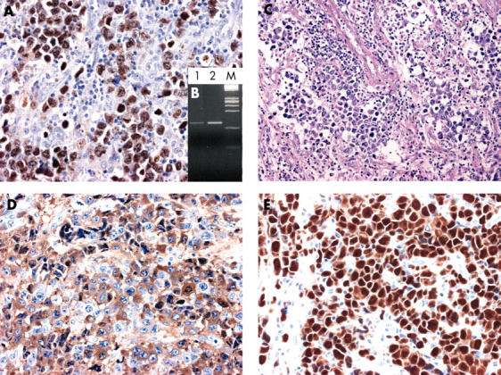
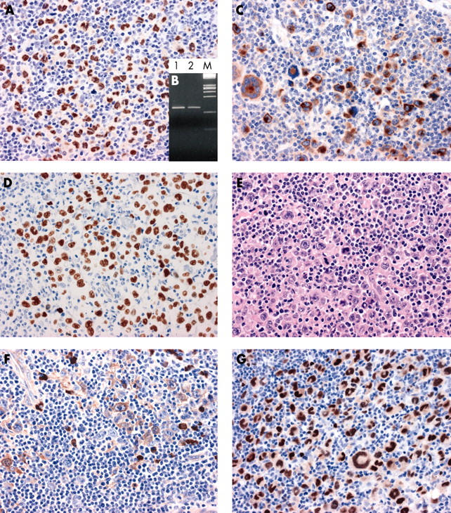
Methods/results: This paper provides evidence for the existence of a previously undescribed KSHV/HHV-8 associated lymphoma in HIV seronegative patients without serous effusions. These lymphomas exhibit a predilection for the lymph nodes and display anaplastic large cell morphology. These tumours were completely devoid of common cell type specific antigens, including epithelial and melanocytic cell markers. B and T cell associated antigens and other commonly used lymphoid markers were absent or weakly demonstrable in a fraction of the tumour cells. Conversely, immunohistochemical studies showed strong immunostaining with plasma cell reactive antibodies.
Conclusions: Analysis of viral infection and immunohistological studies are of primary importance to define this lymph node based KSHV/HHV-8 associated lymphoma with anaplastic large cell morphology and plasmablastic immunophenotype occurring in HIV seronegative patients without serous effusions.
Figures
Similar articles
-
Human Herpesvirus Type 8-associated Large B-cell Lymphoma: A Nonserous Extracavitary Variant of Primary Effusion Lymphoma in an HIV-infected Man: A Case Report and Review of the Literature.Clin Lymphoma Myeloma Leuk. 2016 Jun;16(6):311-21. doi: 10.1016/j.clml.2016.03.013. Epub 2016 Apr 1. Clin Lymphoma Myeloma Leuk. 2016. PMID: 27234438 Free PMC article. Review.
-
High incidence of Kaposi sarcoma-associated herpesvirus infection in HIV-related solid immunoblastic/plasmablastic diffuse large B-cell lymphoma.Leukemia. 2005 May;19(5):851-5. doi: 10.1038/sj.leu.2403709. Leukemia. 2005. PMID: 15744337
-
Extracavitary KSHV-associated large B-Cell lymphoma: a distinct entity or a subtype of primary effusion lymphoma? Study of 9 cases and review of an additional 43 cases.Am J Surg Pathol. 2012 Aug;36(8):1129-40. doi: 10.1097/PAS.0b013e31825b38ec. Am J Surg Pathol. 2012. PMID: 22790853
-
HHV-8-associated lymphoma: state-of-the-art review.Acta Haematol. 2007;117(3):129-31. doi: 10.1159/000097459. Epub 2006 Nov 29. Acta Haematol. 2007. PMID: 17135725 Review.
-
Human herpesvirus 8-associated solid lymphomas that occur in AIDS patients take anaplastic large cell morphology.Mod Pathol. 2000 Jan;13(1):77-85. doi: 10.1038/modpathol.3880012. Mod Pathol. 2000. PMID: 10658913
Cited by
-
KSHV/HHV8-positive large B-cell lymphomas and associated diseases: a heterogeneous group of lymphoproliferative processes with significant clinicopathological overlap.Mod Pathol. 2020 Jan;33(1):18-28. doi: 10.1038/s41379-019-0365-y. Epub 2019 Sep 16. Mod Pathol. 2020. PMID: 31527708 Review.
-
Extracavitary KSHV-positive solid lymphoma: a large B-cell lymphoma within the spectrum of primary effusion lymphoma.Am J Surg Pathol. 2013 Sep;37(9):1460-1. doi: 10.1097/PAS.0b013e31829caada. Am J Surg Pathol. 2013. PMID: 24076781 Free PMC article. No abstract available.
-
Human Herpesvirus Type 8-associated Large B-cell Lymphoma: A Nonserous Extracavitary Variant of Primary Effusion Lymphoma in an HIV-infected Man: A Case Report and Review of the Literature.Clin Lymphoma Myeloma Leuk. 2016 Jun;16(6):311-21. doi: 10.1016/j.clml.2016.03.013. Epub 2016 Apr 1. Clin Lymphoma Myeloma Leuk. 2016. PMID: 27234438 Free PMC article. Review.
-
Lymph node-based disease and HHV-8/KSHV infection in HIV seronegative patients: report of three new cases of a heterogeneous group of diseases.Int J Hematol. 2011 Jun;93(6):795-801. doi: 10.1007/s12185-011-0849-0. Epub 2011 Apr 21. Int J Hematol. 2011. PMID: 21509436
-
Kaposi sarcoma-associated herpesvirus/human herpesvirus 8 and lymphoproliferative disorders.J Clin Pathol. 2007 Dec;60(12):1350-7. doi: 10.1136/jcp.2007.047969. J Clin Pathol. 2007. PMID: 18042691 Free PMC article. Review.
References
-
- Chang Y, Cesarman E, Pessin MS, et al. Identification of herpesvirus-like DNA sequences in AIDS-associated Kaposi’s sarcoma. Science 1994;226:1865–9. - PubMed
-
- Cesarman E, Knowles DM. The role of Kaposi’s sarcoma-associated herpesvirus (KSHV/HHV-8) in lymphoproliferative diseases. Semin Cancer Biol 1999;9:165–74. - PubMed
-
- Cesarman E, Chang Y, Moore PS, et al. Kaposi’s sarcoma-associated herpesvirus-like DNA sequences in AIDS-related body cavity-based lymphomas. N Engl J Med 1995;332:1186–91. - PubMed
-
- Soulier J, Grollet L, Oksenhendler E, et al. Kaposi’s sarcoma-associated herpesvirus-like DNA sequences in multicentric Castleman’s disease. Blood 1995;86:1276–80. - PubMed
-
- Dupin N, Diss TL, Kellam P, et al. HHV-8 is associated with a plasmablastic variant of Castleman disease that is linked to HHV-8-positive plasmablastic lymphoma. Blood 2000;95:1406–12. - PubMed
Publication types
MeSH terms
Substances
LinkOut - more resources
Full Text Sources