

A practical approach to the clinical diagnosis of Ewing's sarcoma/primitive neuroectodermal tumour and other small round cell tumours sharing EWS rearrangement using new fluorescence in situ hybridisation probes for EWSR1 on formalin fixed, paraffin wax embedded tissue
- PMID: 16189150
- PMCID: PMC1770737
- DOI: 10.1136/jcp.2004.025502
A practical approach to the clinical diagnosis of Ewing's sarcoma/primitive neuroectodermal tumour and other small round cell tumours sharing EWS rearrangement using new fluorescence in situ hybridisation probes for EWSR1 on formalin fixed, paraffin wax embedded tissue
Abstract
Background: Over 90% of Ewing's sarcoma/primitive neuroectodermal tumour (ES/PNET) cases have the t(11;22) chromosomal rearrangement, which is also found in other small round cell tumours, including desmoplastic small round cell tumour (DSRCT) and clear cell sarcoma (CCS). Although this rearrangement can be analysed by fluorescence in situ hybridisation (FISH) using routinely formalin fixed, paraffin wax embedded (FFPE) tissues when fresh or frozen tissues are not available, a sensitive and convenient detection method is needed for routine clinical diagnosis.
Aims: To investigate the usefulness of newly developed probes for detecting EWS rearrangement resulting from chromosomal translocations using FISH and FFPE tissue in the clinical diagnosis of ES/PNET, DSRCT, and CCS.
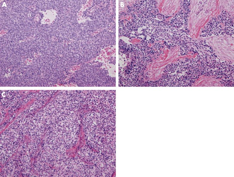
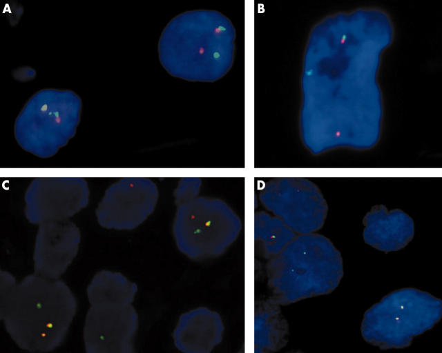
Methods: Sixteen ES/PNETs, six DSRCTs, and six CCSs were studied. Three poorly differentiated synovial sarcomas, three alveolar rhabdomyosarcomas, and three neuroblastomas served as negative controls. Interphase FISH analysis was performed on FFPE tissue sections with a commercially available EWSR1 (22q12) dual colour, breakapart rearrangement probe.
Results: One fused signal and one split signal of orange and green, demonstrating rearrangement of the EWS gene, was detected in 14 of 16 ES/PNETs, all six DRSCTs, and five of six CCSs, but not in the negative controls.
Conclusions: Interphase FISH using this newly developed probe is sensitive and specific for detecting the EWS gene on FFPE tissues and is of value in the routine clinical diagnosis of ES/PNET, DSRCT, and CCS.
Figures
References
-
- Aurias A, Rimbaut C, Buffe D, et al. Translocation involving chromosome 22 in Ewing’s sarcoma. A cytogenetic study of four fresh tumors. Cancer Genet Cytogenet 1984;12:21–5. - PubMed
-
- Turc-Carel C, Philip I, Berger MP, et al. Chromosomal study of Ewing’s sarcoma (ES) cell lines. Consistency of a reciprocal translocation t(11;22)(q24;q12). Cancer Genet Cytogenet 1984;12:1–19. - PubMed
Publication types
MeSH terms
Substances
LinkOut - more resources
Full Text Sources
Other Literature Sources
Medical