

Deletion of the core region of 5' HS2 of the mouse beta-globin locus control region reveals a distinct effect in comparison with human beta-globin transgenes
- PMID: 16189270
- PMCID: PMC1895626
- DOI: 10.1182/blood-2005-06-2308
Deletion of the core region of 5' HS2 of the mouse beta-globin locus control region reveals a distinct effect in comparison with human beta-globin transgenes
Abstract
The beta-globin locus control region (LCR) is a large DNA element that is required for high-level expression of beta-like globin genes from the endogenous mouse locus or in transgenic mice carrying the human beta-globin locus. The LCR encompasses 6 DNaseI hypersensitive sites (HSs) that bind transcription factors. These HSs each contain a core of a few hundred base pairs (bp) that has most of the functional activity and exhibits high interspecies sequence homology. Adjoining the cores are 500- to 1000-bp "flanks" with weaker functional activity and lower interspecies homology. Studies of human beta-globin transgenes and of the endogenous murine locus show that deletion of an entire HS (core plus flanks) moderately suppresses expression. However, human transgenes in which only individual HS core regions were deleted showed drastic loss of expression accompanied by changes in chromatin structure. To address these disparate results, we have deleted the core region of 5'HS2 from the endogenous murine beta-LCR. The phenotype was similar to that of the larger 5'HS2 deletion, with no apparent disruption of chromatin structure. These results demonstrate that the greater severity of HS core deletions in comparison to full HS deletions is not a general property of the beta-LCR.
Figures


 , the CMV-HyTK gene after homologous recombination. The expected band sizes are noted. (B) ΔHS2 full deletion Southern blot. Lanes 1 and 5, ΔHS2 full/S; lane 2, D/S; and lanes 3 and 4, HS2(HR)/S. (C) ΔHS2 core deletion Southern blot. Lanes 1 and 3, ΔHS2c/S; lane 2, ΔHS2c(inv)/S; lane 4, HS2(HR)/S; and lane 5, D/S.
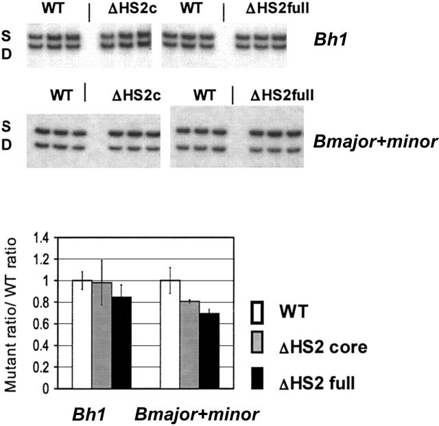


References
-
- Wang Y, Macke J, Merbs S, et al. A locus control region adjacent to the human red and green visual pigment genes. Neuron. 1992;9: 429-440. - PubMed
-
- Madisen L, Groudine M. Identification of a locus control region in the immunoglobulin heavy-chain locus that deregulates c-myc expression in plasmacytoma and Burkitt's lymphoma cells. Genes Dev. 1994;8: 2212-2226. - PubMed
-
- Stamatoyannopoulos G, Grosveld F. Hemoglobin switching. In: Stamatoyannopoulos G, Majerus PW, Perlmutter RM, Varmus H, eds. The Molecular Basis of Blood diseases. Philadelphia, PA: Saunders; 2001: 135-182.
Publication types
MeSH terms
Substances
Grants and funding
LinkOut - more resources
Full Text Sources
Molecular Biology Databases
