. 2006 Jan 15;107(2):483-91.
doi: 10.1182/blood-2005-05-2133. Epub 2005 Sep 27.

Stable gene transfer and expression in human primary T cells by the Sleeping Beauty transposon system

Affiliations

Stable gene transfer and expression in human primary T cells by the Sleeping Beauty transposon system

Xin Huang et al. Blood. .

Abstract

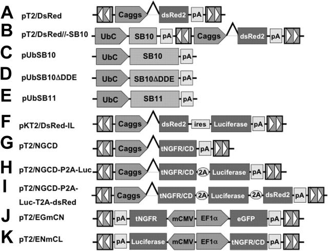
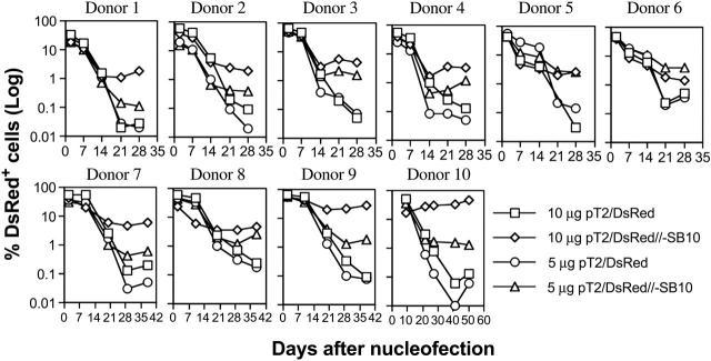
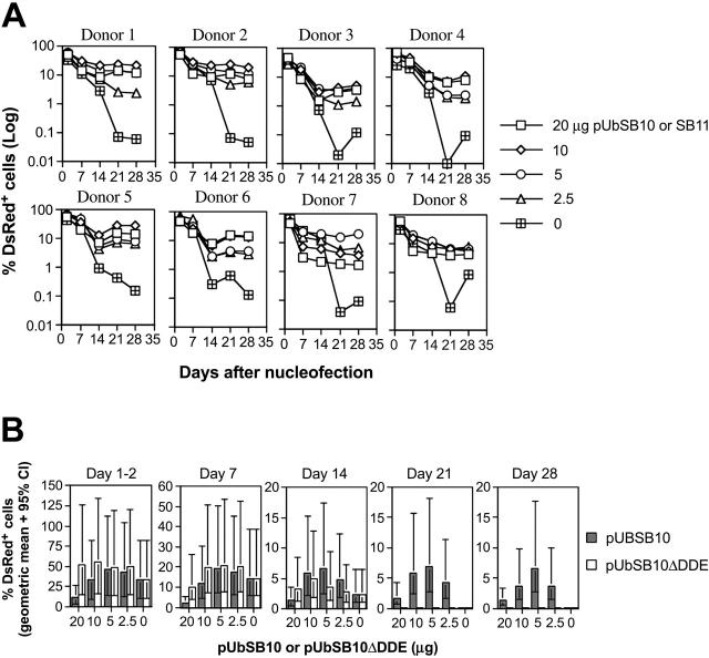
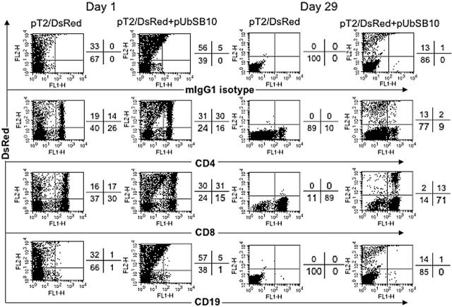
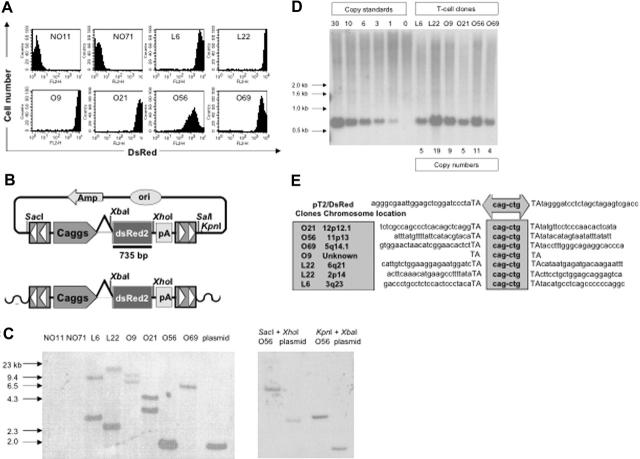
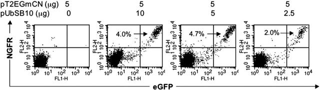
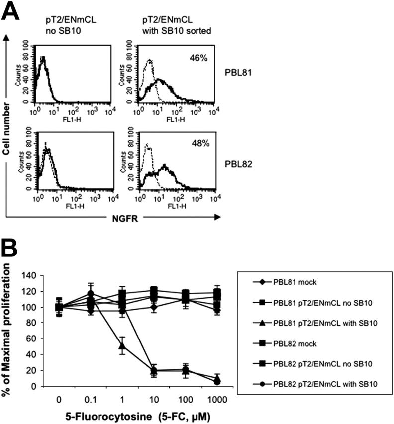
The Sleeping Beauty (SB) transposon system is a nonviral DNA delivery system in which a transposase directs integration of an SB transposon into TA-dinucleotide sites in the genome. To determine whether the SB transposon system can mediate stable gene expression in human T cells, primary peripheral blood lymphocytes (PBLs) were nucleofected with SB vectors carrying a DsRed reporter gene. Plasmids containing the SB transposase on the same molecule as (cis) or on a molecule separate from (trans) the SB transposon mediated long-term and stable reporter gene expression in human primary T cells. Sequencing of transposon:chromosome junctions confirmed that stable gene expression was due to SB-mediated transposition. In other studies, PBLs were successfully transfected using the SB transposon system and shown to stably express a fusion protein consisting of (1) a surface receptor useful for positive T-cell selection and (2) a "suicide" gene useful for elimination of transfected T cells after chemotherapy. This study is the first report demonstrating that the SB transposon system can mediate stable gene transfer in human primary PBLs, which may be advantageous for T-cell-based gene therapies.


Figures

Figure 1.
Figure 1.
The Sleeping Beauty (SB) transposon vectors used in this study. The SB transposon system consists of 2 components: the inverted repeat/direct repeats (IR/DR, indicated by arrowheads) flanking the gene of interest, and the expression cassette encoding the transposase. Caggs is a chimeric promoter derived from chicken β-actin and cytomegalovirus immediate-early promoter sequences. UbC indicates human ubiquitin C promoter; SB10, transposase; SB10ΔDDE, inactive transposase due to deletion of the catalytic domain; tNGFR/CD, truncated human nerve growth factor receptor and cytosine deaminase fusion gene; P2A and T2A, “self-cleaving” 2A peptides derived from porcine teschovirus-1 and Thosea asigna virus have been shown to express multiple genes. pT2/EGmCN and pT2/ENmCL were bidirectional SB vectors in which synthetic bidirectional promoters were used. mCMV indicates minimal CMV promoter; EF1α, human elongation factor 1α promoter; IRES, internal ribosomal entry sites; DsRed2, red fluorescent protein; Luciferase, bioluminescent reporter gene; and pA, polyadenylation signal.
Figure 2.
Figure 2.
Stable transgene expression in human primary PBLs by the SB transposon cis construct. Freshly isolated PBLs (5 × 10⁶) were nucleofected at concentrations of 10 or 5 μg trans vector pT2/DsRed or cis vector pT2/DsRed//-SB10 using the T-cell Nucleofector kit (Amaxa, Gaithersburg, MD). On day 1 or 2 after nucleofection, a fraction of cells was analyzed for DsRed expression by flow cytometry, and the remaining cells were stimulated with anti-CD3/28 beads for 3 to 5 days. After removal of beads, the cells were cultured in human T-cell medium with IL-2 (50 IU/mL) and IL-7 (10 ng/mL). DsRed transgene expression was analyzed at the indicated time points by flow cytometry. The cells were restimulated once every 10 to 14 days. The data from 10 individual PBLs are shown.
Figure 3.
Figure 3.
Stable transgene expression in human primary PBLs by the SB transposon trans construct and functional transposase. (A) SB trans delivery. Freshly isolated PBLs (5 × 10⁶) were co-nucleofected with 5 μg of pT2/DsRed and different amounts of pUbSB10 or pUbSB11 (donors 7 and 8 only) (20, 10, 5, 2.5, and 0 μg). DsRed expression was analyzed on days 1, 2, 7, 14, 21, and 28. The data from 8 individual PBLs are shown. (B) Comparison of wild-type and mutant SB10 in trans delivery. Columns represent geometric means with 95% confidence intervals of 6 independent experiments. P values to compare SB10 and SB10ΔDDE on days 21 and 28 were all less than .001.
Figure 4.
Figure 4.
Immunophenotyping of transfected T cells. PBLs from 2 donors were nucleofected with pT2/DsRed or with pT2/DsRed plus pUbSB10, or without DNA (mock, data not shown), and then the cells were stained with anti-CD4, CD8, CD19, and isotype mouse IgG1 mAbs on days 1, 15 (not shown), and 29 for analysis by flow cytometry. Representative data from one donor is shown. Similar data from at least 3 other donors were obtained 3 to 4 weeks after transfection (not shown).
Figure 5.
Figure 5.
Molecular analyses of transposition in human T cells. (A) DsRed transgene expression in transposed T-cell clones 4 months after gene transfer. (B) Schematic representation of a DsRed probe used in this study. Top row shows the circular transposon-encoded plasmid nucleofected into T cells and the location of the 735-bp DsRed probe. Bottom row shows the loss of flanking restriction enzyme recognition sequences after transposase-mediated integration into genomic DNA (wavy line). (C) Integration assay by Southern blotting. Southern blot of genomic DNA digested with SalI and XbaI and hybridized with a DsRed probe. The SalI site located outside the IR/DR is lost, resulting in the absence of a detectable 1.7-kb plasmid-specific band. Southern blot of genomic DNA from clone O56 digested with SacI and XhoI as well as KpnI and XbaI and hybridized with the same probe. The SacI and KpnI sites flanking IR/DRs are lost, resulting in the absence of detectable 2.8- and 1.7-kb plasmid-specific bands, respectively. (D) Determination of per-cell copy numbers by Southern blot. A 1-copy standard of pT2/DsRed was equal to 11 pg. Copy standards were mixed with 10 μg genomic DNA isolated from bulk activated T cells. Transgene copy numbers for each T-cell clone were calculated from the intensities of the bands spanning the DsRed gene in comparison to a copy number standard after linear regression analysis. Band intensities were quantified using a PhosphorImager. (E) Transposition assay. Transposon insertion site sequences were determined using a linker-mediated PCR technique described in “Materials and methods.” Plasmid backbone sequences of pT2/DsRed are shown. Duplicated TA dinucleotide target site is in capital letters. Transposon-specific sequences are in the center box. Amplified transposon:chromosome junction sequences were subjected to BlastN analysis against the human genome using the ENSEMBL database (chromosome locations indicated in shaded box on the left). Genome-specific primers were designed in accordance with identified chromosome positions to amplify the opposing flanking sequence and confirm insertion into a single TA dinucleotide.
Figure 6.
Figure 6.
Dual gene expression in human T cells. PBLs from 2 donors were nucleofected with the SB dual gene plasmid pT2/EGmCN (5 μg) ± pUbSB10 (10, 5, 2.5, and 0 μg). The cells were activated by anti-CD3/28 beads on day 2 and assayed for eGFP and NGFR expression on days 7 (data not shown) and 21. Similar data were also obtained from another donor.
Figure 7.
Figure 7.
Functional therapeutic gene expression in human T cells by SB-mediated transposition. (A) NGFR expression in the SB-nucleofected T cells (solid line). A murine IgG1 isotype mAb was used (broken line). (B) Cell killing by 5-FC. Human T cells nucleofected with SB transposon DNA (2 × 10⁵ cells/well) or CEM cells (104 cells/well) transduced with retrovirus expressing NGCD were exposed to different concentrations of 5-fluorocytosine (5-FC, Sigma) in 96-well flat-bottom plates for 5 days. A colorimetric assay kit (CellTiter 96, Promega) based on the conversion of a tetrazolium dye to formazan was used to quantitate the number of viable cells on day 5. Absorbance was measured at 565 nm using a microplate spectrophotometer (Bio-Tek Instruments, Winooski, VT). CEM cells alone or transduced with retrovirus expressing NGCD were used as negative and positive controls (data not shown). The data represent mean and standard deviation of 10 replicates. This experiment was repeated twice with similar results. Similar results also were obtained with the plasmid pT2/NGCD (data not shown).

References

    1. Sadelain M, Riviere I, Brentjens R. Targeting tumours with genetically enhanced T lymphocytes. Nat Rev Cancer. 2003;3: 35-45. - PubMed
    1. Rossig Cl, Brenner M. Genetic modification of T lymphocytes for adoptive immunotherapy. Mol Ther. 2004;10: 5-18. - PubMed
    1. Tiberghien P. “Suicide” gene for the control of graft-versus-host disease. Curr Opin Hematol. 1998;5: 478-482. - PubMed
    1. Greenberg PD, Riddell SR. Deficient cellular immunity-finding and fixing the defects. Science. 1999;285: 546-551. - PubMed
    1. Fisher A, Hacein-Bey S, Cavazzana-Calvo M. Gene therapy of severe combined immunodeficiencies. Nat Rev Immunol. 2002;2: 615-621. - PubMed

Publication types

Substances