. 2006 Jan 15;107(2):689-97.
doi: 10.1182/blood-2005-05-2125. Epub 2005 Sep 27.

Ablation of oncogenic ALK is a viable therapeutic approach for anaplastic large-cell lymphomas

Affiliations

Ablation of oncogenic ALK is a viable therapeutic approach for anaplastic large-cell lymphomas

Roberto Piva et al. Blood. .

Abstract

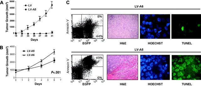
Anaplastic large-cell lymphomas (ALCLs) carry chromosome translocations in which the anaplastic lymphoma kinase (ALK) gene is fused to several partners, most frequently, the NPM1 gene. We have demonstrated that the constitutive activation of ALK fusion proteins results in cellular transformation and lymphoid neoplasia. Herein, we specifically down-regulated ALK protein expression by using small hairpin RNA (shRNA) targeting a sequence coding for the catalytic domain of ALK. The ablation of ALK leads to the down-modulation of known ALK downstream effectors, cell growth arrest, and reversion of the transformed phenotype of ALK(+) mouse embryonic fibroblasts in vitro and in vivo. In human ALCL cells lentiviral-mediated ALK knock-down leads to G(1) cell-cycle arrest and apoptosis in vitro and tumor growth inhibition and regression in vivo. Using a specific approach we have demonstrated that the survival and growth of ALK(+) ALCLs are strictly dependent on ALK activation and signaling. Therefore, ALK is a viable target for therapeutic intervention and its inactivation might represent a pivotal approach for the treatment of ALK lymphomas and other ALK-dependent human tumors.


Figures

Figure 1.
Figure 1.
Selection of ALK shRNA. (A) ALK-A5 efficiently inhibits NPM-ALK protein expression. HEK-293T cells were cotransfected with Pallino NPM-ALK (0.2 μg) and 1 of 6 different pSUPER vectors carrying shRNA specific for ALK sequences (pS-A1-6; 0.8 μg). Untransfected cells were also included (–). Lysates (72 hours) were immunoblotted using a specific anti-ALK antibody recognizing the cytoplasmic region of the ALK-R. Protein loading was normalized using anti–α-tubulin antibody. (B) shRNA ALK-A5 does not modulate the expression of TPR-MET. HEK-293T cells transfected with TRP-MET and ALK-A5 pSUPER vectors express unmodified levels of TPR-MET. However, TPR-MET expression is down-modulated after cotransfection with a specific TPR-MET (TM2) shRNA construct (top panel). HEK-293T cells transfected NPM-ALK and the TPR-MET shRNA cassette expressed unmodified levels of NPM-ALK (bottom panel). (C) ALK5 inhibits ATIC-ALK protein expression. HEK-293T cells were cotransfected with Pallino ATIC-ALK and ALK-pSUPER shRNA interfering sequences. Expression of ATIC-ALK protein was determined by Western blot analysis as described in “Materials and methods.” (D) ALK-A5 inhibits ALK-R protein expression. HEK-293T cells were cotransfected with Pallino ALK-R and pSUPER-ALK shRNA interfering sequences. Protein loading was normalized using a rabbit polyclonal antibody to CDK2.
Figure 2.
Figure 2.
ALK siRNA reverts NPM-ALK–mediated transformation of MEF cells in vitro and in vivo. (A) Suppression of NPM-ALK expression leads to down-regulation of known downstream targets of ALK. Lysates from NPM-ALK Tet-Off MEFs infected with pSRG-A5 virus (semiconfluent cells in absence of doxycycline) before (40% EGFP+) or after (> 90% EGFP+) puromycin selection were immunoblotted with the indicated antibodies. (B) NPM-ALK MEF cells lose their transformed phenotype in absence of NPM-ALK expression. Cell cultures in absence of doxycyline are rescued from the ALK-mediated transformed phenotype by the expression of ALK-A5 shRNA (contrast phase microscopy, left panels). Control and pSRG-A5–infected NPM-ALK Tet-Off MEF cells were stained with anti-ALK antibodies, followed by biotin-conjugated horse antimouse antibody and streptavidin-Cy3. Cells were counterstained with Hoechst 33258 and visualized with a Leica fluorescence microscope using a 63× objective (right panels). (C) pSRG-A5 expression prevents NPM-ALK MEF cell growth in immunocompromised mice. NPM-ALK Tet-Off MEF cells transduced with pSRG-A5 or control pSRG were first selected with puromycin (> 90% EGFP+), inoculated (106 cells/mouse) subcutaneously into athymic Nu/Nu mice recipients (3 mice for each construct). • indicates pSRG; ▪, pSRG-A5. Tumor growth was monitored over time. These findings are representative of 2 experiments. Error bars indicate SD.
Figure 3.
Figure 3.
ALK shRNA inhibits the growth of human ALCL cells. (A) Percentages of human (TS, 80%) and murine (CD4-43, 8%) ALK+ cells transduced with pSRG-A5 vector as measured by EGFP expression by FACS analysis. (B-F) Human and murine ALCL ALK-A5+ cells decrease over time. Percentages of EGFP+ cells in pSRG-A5 or pSRG retrovirally transduced human ALCL cells SU-DHL-1, TS, and Karpas 299, human lymphoblastoid cells CCRF-CEM (ALK), and murine NPM-ALK (CD4-43) cells were determined over time. These findings are representative of at least 3 different experiments. In B-F, ♦ indicates pSRG-A6; ▪, pSRG-A5. (G) Percentages of BrdU/ALK-A5+ ALCL cells decrease over time. ALK-A5–infected TS cells were cultured over time and the percentages of BrdU+ cells within EGFP and + cells were calculated at different intervals. These findings are representative of at least 3 different experiments. Numbers represent the percentages of positive cells for each quadrant. (H) Percentages of BrdU+ TS cells over time after transduction with pSRG-A5 and pSRG-A6 viruses. TS cells were transduced with retroviral ALK-A5 or ALK-A6 pSRG vectors. BrdU incorporation of EGFP+ and EGFP cells was established as described in “Materials and methods.” ▪ indicates pSRG-A5; □, pSRG-A6. Percentages of total are indicated. Normalized ratio of BrdU+ cells among EGFP+ or EGFP cells was calculated over time. (A-F, H) Error bars indicate SD.
Figure 4.
Figure 4.
Growth of ALK-A5 retrovirally infected cells is inhibited in presence of low FCS concentration or cytotoxic agents. (A) ALK-A5 or ALK-A6 pSRG– or pSRG-infected cells were cultured in the presence of different concentrations of FCS. The percentages of EGFP+ cells were calculated over time. Data are reported as ratios of ALK-A5 versus ALK-A6 pSRG EGFP+ cells. (B) Retrovirally infected cells (3 days after transduction) were cultured with different concentration of bleomycin (B) or cyclophosphamide (C). Percentages of EGFP+ cells were obtained by flow cytometry and ratios between ALK-A5 versus ALK-A6 pSRG EGFP+ cells were calculated. In A-C, ▪ represents pSRG-A5; □, pSRG-A6.
Figure 5.
Figure 5.
Lentiviral ALK shRNA abrogates NPM-ALK expression and signaling. (A) Lentivirus infection with ALK-A5 leads to an efficient down-regulation of NPM-ALK protein expression. TS cells were transduced with ALK sh-RNA lentivirus (300 μL) and then harvested to determine the NPM-ALK protein expression over time. Immunoblotting with anti-GFP antibodies was used to check the expression of the report gene within the lentiviral cassette. (B). Loss of NPM-ALK is specifically observed in cells infected with ALK-A5. TS cells were transduced with ALK-A5 or TM2 viruses, cultured for 96 hours, and then stained with anti-ALK antibody. Immune complexes were visualized using biotin-conjugated rabbit antimouse antibody followed by streptavidin-Cy3 (right panels). Nuclei were identified using Hoechst 33258 (left panels). EGFP expression is shown in middle panels. Objective magnification, 40×. (C) TS cells transduced with ALK-A5 show phospho-Stat3, phospho-AKT, and Jun B down-modulation. EV indicates empty vector.
Figure 6.
Figure 6.
Lentiviral ALK shRNA leads to cell-cycle arrest and apoptosis of ALCL cells. (A-B) Anti–ALK-A5 leads to G1 cell-cycle arrest. TS cells transduced with the indicated constructs were harvested 96 hours after infection and stained with propidium iodide (PI) to determine the corresponding fractions in G0/G1, S, and G2-M phases. Lysates were analyzed by Western blotting with the indicated antibodies. (C) Anti–ALK-A5 leads to cell death. Percentages of TMRM cells were calculated in TS cells 4 and 5 days after infection. (A, C) Error bars indicate SD. (D) Loss of NPM-ALK leads to caspase activation. TS cells transduced with empty vector, TM2, or ALK were harvested 96 hours after transduction and immunoblotted with the indicated antibodies. These findings are representative of 4 different experiments.
Figure 7.
Figure 7.
NPM-ALK silencing impairs the growth of human ALK+ cells in vivo. (A) TS cells transduced with ALK-A5 do not generate xenograph tumors in immunocompromised animals. TS cells transduced with ALK-A5 or with the control ALK-A6 lentivirus were injected into FoxChase (C.B-17) SCID mice 48 hours after infection. Tumor growth was observed over time. A total of 12 mice (6 for each group) was studied. (B) ALCL tumor mass growth is hindered by the intratumoral injection of lentiviruses carrying the ALK-A5 shRNA. FoxChase (C.B-17) SCID mice injected with TS cells (2 × 10⁶) were treated intratumorally when the tumor masses were about 1 cm3 with 50μL (0.5-1 × 10⁸ TU) of concentrated lentivirus preparations carrying ALK-A5 or control ALK-A6 shRNA, every alternative day, 3 times. Tumor growth was determined over time. (A-B) Error bars indicate SD. (C) ALK-A5 shRNA leads to cell death in vivo. After treatment, tumor cells were isolated and stained with PE–annexin V and evaluated by flow cytometry. Percentages of EGFP+ cells are indicated (left panels). Representative histologic sections (hematoxylin and eosin [H&E]) of tumor masses derived from animals treated with LV-A6 or LV-A5 preparations are shown as indicated (second panels from left; objective magnification, 20×). The same paraffin-embedded tissue sections were also stained for DNA breaks with a TUNEL method. Hoechst counterstain identifies the nuclei (third panels from left; objective magnification, 40×). DNA break points are demonstrated by the positive FITC stains (fourth panels from left; objective magnification, 40×). Variable percentages of positive cells were observed in different areas within the tumor cells treated with ALK-A5 lentiviral preparations. These findings are representative of 3 different experiments with a total of 12 mice for each group.

References

    1. Stein H, Foss HD, Durkop H, et al. CD30(+) anaplastic large cell lymphoma: a review of its histopathologic, genetic, and clinical features. Blood. 2000;96: 3681-3695. - PubMed
    1. Morris SW, Kirstein MN, Valentine MB, et al. Fusion of a kinase gene, ALK, to a nucleolar protein gene, NPM, in non-Hodgkin's lymphoma. Science. 1994;263: 1281-1284. - PubMed
    1. Duyster J, Bai RY, Morris SW. Translocations involving anaplastic lymphoma kinase (ALK). Oncogene. 2001;20: 5623-5637. - PubMed
    1. Cessna MH, Zhou H, Sanger WG, et al. Expression of ALK1 and p80 in inflammatory myofibroblastic tumor and its mesenchymal mimics: a study of 135 cases. Mod Pathol. 2002;15: 931-938. - PubMed
    1. Onciu M, Behm FG, Downing JR, et al. ALK-positive plasmablastic B-cell lymphoma with expression of the NPM-ALK fusion transcript: report of 2 cases. Blood. 2003;102: 2642-2644. - PubMed

Publication types

MeSH terms