Review
. 2005 Nov;6(11):967-77.
doi: 10.1111/j.1600-0854.2005.00339.x.

Wrapping things up about virus RNA replication

Affiliations
Review

Wrapping things up about virus RNA replication

Jason Mackenzie. Traffic. 2005 Nov.

Abstract

All single-stranded 'positive-sense' RNA viruses that infect mammalian, insect or plant cells rearrange internal cellular membranes to provide an environment facilitating virus replication. A striking feature of these unique membrane structures is the induction of 70-100 nm vesicles (either free within the cytoplasm, associated with other induced vesicles or bound within a surrounding membrane) harbouring the viral replication complex (RC). Although similar in appearance, the cellular composition of these vesicles appears to vary for different viruses, implying different organelle origins for the intracellular sites of viral RNA replication. Genetic analysis has revealed that induction of these membrane structures can be attributed to a particular viral gene product, usually a non-structural protein. This review will highlight our current knowledge of the formation and composition of virus RCs and describe some of the similarities and differences in RNA-membrane interactions observed between the virus families Flaviviridae and Picornaviridae.


Figures

Figure 1
Figure 1
Schematic representation of the Flavivirus genome organization and polyprotein‐processing events associated with replication. Cleavage sites depicted with a ▾ represent cleavage by host‐signal peptidase within the lumen of the endoplasmic reticulum. Sites represented with a formula image 
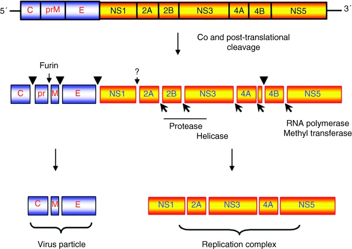
depict cleavage by the viral‐encoded protease NS3 with cofactor NS2B. The cleavage between NS1 and NS2A is currently not well understood but is performed by a host‐cell protease in the lumen of the endoplasmic recticulum.
Figure 2
Figure 2
Ultrastructural observations of Kunjin virus‐infected Vero cells at 24 h post‐infection. Panel A reveals immunogold labelling of a thawed cryosection prepared from Kunjin virus‐infected cells with antibodies raised against protein disulphide isomerase (PDI) and 10 nm protein‐A gold. Note the labelling of the lumen of the rER continuous with the convoluted membranes (CM) but no labelling within this structure. Adjacent vesicle packets (VPs) are also devoid of the PDI antibodies. Magnification bar represents 200 nm. Schematic representation of the flavivirus life cycle is depicted in PanelB.Step 1 proposes the incorporation of cytoplasmic viral RNA into the VP for replication by the replication complex. (2) The transcribed viral RNA is then transported to the CM/PC for translation and proteolytic cleavage via both host‐ and viral‐encoded proteases. (3) Subsequently, the RNA is encapsidated by the core protein, and virion assembly proceeds via budding into the ER lumen. (4) Virion maturation occurs via transit through the Golgi apparatus before release through the plasma membrane (PM) via exocytosis (5). Redistribution of trans‐Golgi network (TGN) and intermediate compartment (IC) host proteins to the VP and CM/paracrystalline arrays (PC), respectively, are highlighted.
Figure 3
Figure 3
Schematic representation of the genomic organization of the Picornavirus genome and polyprotein species generated via virus‐specific proteolysis. Many of these polyprotein species have specific roles during replication of picornaviruses.
Figure 4
Figure 4
Ultrastructural analysis of Parechovirus‐infected BSC‐1 cells at 6 h post‐infection. In (A), the resin‐embedded section reveals vesicle clusters (VCs) accumulating in the perinuclear region. The vesicles range in size from approximately 100–800 nm. B) immunogold labelling of cryosections from parechovirus‐infected cells reveals the vesicle clusters heavily decorated with antibodies to dsRNA and 10 nm protein‐A gold.C)Schematic representation of membrane rearrangements observed during picornavirus infection. Anterograde transport carriers are restricted at different stages of the secretory pathway depending on the different viruses investigated. This restriction in transport of protein and membrane to the Golgi apparatus leads to the eventual disintegration of the Golgi due to the redistribution of Golgi proteins and membrane to the ER via retrograde movement. Nu, nucleus. Magnification bars represent 1 µm and 200 nm in Panels A and B, respectively.
Figure 5
Figure 5
Immunofluorescene analysis of EMCV‐infected (A) and Echovirus 11‐infected (B) BSC‐1 cells at 6 h post‐infection. Co‐localization of anti‐dsRNA (labelled with Alexa Fluor 488; green) and anti‐ERGIC53 (labelled with Alexa Fluor 568; red) antibodies is observed within discrete cytoplasmic foci representing the picornavirus replication complex (highlighted with arrowheads). Dual localization is recognized as a yellow hue in both panels.

References

    1. Egger D, Wolk B, Gosert R, Bianchi L, Blum HE, Moradpour D, Bienz K. Expression of hepatitis C virus proteins induces distinct membrane alterations including a candidate viral replication complex. J Virol 2002;76: 5974–5984. - PMC - PubMed
    1. Bienz K, Egger D, Pasamontes L. Association of polioviral proteins of the P2 genomic region with the viral replication complex and virus‐induced membrane synthesis as visualized by electron microscopic immunocytochemistry and autoradiography. Virology 1987;160: 220–226. - PubMed
    1. Gazina EV, Mackenzie JM, Gorrell RJ, Anderson DA. Differential requirements for COPI coats in formation of replication complexes among three genera of Picornaviridae. J Virol 2002;76: 11113–11122. - PMC - PubMed
    1. Kujala P, Ikaheimonen A, Ehsani N, Vihinen H, Auvinen P, Kaariainen L. Biogenesis of the Semliki Forest virus RNA replication complex. J Virol 2001;75: 3873–3884. - PMC - PubMed
    1. Magliano D, Marshall JA, Bowden DS, Vardaxis N, Meanger J, Lee JY. Rubella virus replication complexes are virus‐modified lysosomes. Virology 1998;240: 57–63. - PubMed

Publication types