. 2005 Oct;86(5):297-307.
doi: 10.1111/j.0959-9673.2005.00439.x.

Evaluation of human knee meniscus biopsies with near-infrared, reflectance confocal microscopy. A pilot study

Affiliations

Evaluation of human knee meniscus biopsies with near-infrared, reflectance confocal microscopy. A pilot study

Vanessa Campo-Ruiz et al. Int J Exp Pathol. 2005 Oct.

Abstract

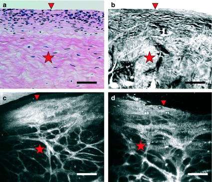
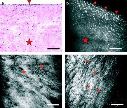
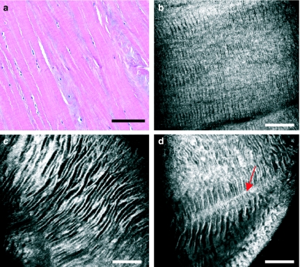
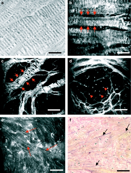
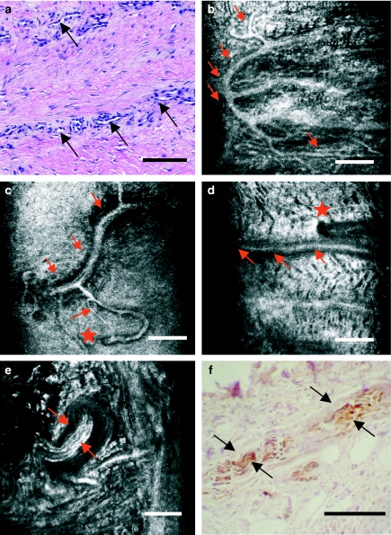
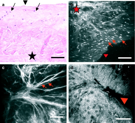
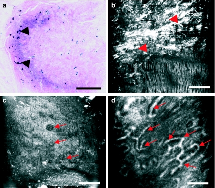
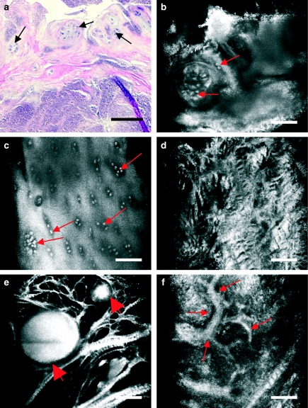
Knee cartilage biopsy is used to confirm the pathology in both clinical and experimental conditions and often guides diagnosis and therapeutic strategies. Current histopathological techniques are time consuming, induce tissue artefacts and often prevent further evaluation, once the tissue has been fixed. Hence, there is a potential need for a fast and nondestructive imaging technique for unfixed tissue. Near-infrared, reflectance confocal microscopy (CM) allows real-time, virtual sectioning of unstained, bulk tissue samples. This pilot study evaluates the use of CM in the assessment of meniscus histopathology in a series of 26 freshly-excised human meniscus samples compared to standard light microscopy of stained sections. CM images of the meniscus show cell and matrix detail, depicting morphologic features of collagen and elastic fibres, vessels and nerve endings. In addition, crystal deposits of gout and pseudogout are also demonstrable. Thus, CM is a novel imaging technique that could enable the pathologist to make a rapid microscopic evaluation of cartilage in a fresh and unfixed fashion.


Figures

Figure 1
Figure 1
Comparison of vertical sections of human knee meniscus as seen with routine haematoxylin – eosin staining examined under light microscopy (a), cross-polarized light microscopy (b) and near-infrared reflectance confocal microscopy imaging of intact tissue exposed to phosphate-buffered saline (b) and to acetic acid (d). The densely cellular top layer (arrowhead) and the more fibrillar interior (star) are readily depicted in the four images. Scale bar represents 100 µm.
Figure 2
Figure 2
Meniscus cells as seen with light microscopy of haematoxylin and eosin-stained sections (a) and with near-infrared, reflectance confocal microscopy (CM) of bulk tissue (b–d). Vertical sections of meniscus (a and b) reveal that cells (arrows) are much more abundant in superficial layers (arrowhead) that in the interior of the meniscus (star). Near the surface, they exhibit an ovoid or fusiform morphology. Virtual sections of meniscus in horizontal planes with CM reveal cells of round morphology (c, arrows) and a surrounding hyperefractile matrix that exhibits fine striae. The location of cells is better seen when tissue is briefly exposed to acetic acid (d, arrows), which renders nuclei more refractive (white in the images). Scale bar represents 100 µm.
Figure 3
Figure 3
Meniscus matrix as seen with light microscopy (a) and with near-infrared, reflectance confocal microscopy (CM) (b–d). With CM, extracellular matrix appears generally hyperefractile, displaying hyporefractile striae that conform a zebra pattern. This zebra pattern is present both in the superficial (b) and in the intermediate-deep layers (c and d) of the meniscus, progressively increasing the width of its striae. Striae appear intersected by perpendicular fibrous tracts of greater refractility (d, arrow). Scale bar represents 100 µm.
Figure 4
Figure 4
Meniscus fibres seen with polarized light microscopy (a), routine light microscopy (f) and near-infrared, reflectance confocal microscopy (CM) (b–e). Circumferential (b, between arrows) and radial fibres (c, between arrows) are readily seen with CM. Both types of fibres exhibit areas of zebra pattern. Brief exposure of the tissue to acetic acid highlights the presence of cell nuclei interspersed within the fibrous mesh (d, arrowheads), which were not clearly seen before acetic acid application. Acetic acid also reveals a third type of fibres in CM images (e, arrows). These thin fibres are orientated in different directions and correlate well with elastic fibres stained by Verhoeff-Van Gieson's method (f, arrows). Scale bar represents 100 µm.
Figure 5
Figure 5
Neurovascular structures in the outer third of human knee meniscus, as seen with light microscopy (a and f) and with near-infrared, reflectance confocal microscopy (CM) (b–e). CM readily depicts the localization of vessels and the distribution of their branches (b–d, arrows). The points in which these vessels penetrate from the outer fringe of the meniscus may also be located with precission (d, star). Meniscal myelinated nerve endings are easily distinguishable for their hyperefractile, striated appearance (e, arrows). Images with CM correlate well with those from companion histology sections stained for PGP 9.5 antibody (f, arrows). Scale bar represents 100 µm.
Figure 6
Figure 6
Human knee meniscus exhibiting a degenerative pattern, imaged with routine light microscopy (a) and with near-infrared, reflectance confocal microscopy (CM) (b–d). This degenerative pattern consists of an impairment in the cellular density and distribution, that is, an absence of the decreasing cell gradient from surface (a and b, arrowhead) to depth (a and b, star) described previously for normal meniscus. Matrix architecture appears greatly disorganized and heteromorphic, with thinned matrix fibre bundles (c, arrows) and occasional sites of meniscal tear (d, arrowhead). Scale bar represents 100 µm.
Figure 7
Figure 7
Human knee meniscus affected by gout imaged with routine light microscopy (a) and near-infrared, reflectance confocal microscopy (CM) (b–d). In CM images, urate crystals are visualized as multiple, highly refractive, needle-shaped structures of heterogeneous distribution (b, arrowheads). Note that these crystals inadvertently disappear when tissue is obtained and preserved in saline serum prior to routine histology processing (a, arrows). The surrounding matrix shows increased refractility and disruption in its architecture, with fibres showing different sizes and orientations. Cells appear hypertrophic and with a greater tendency to form clusters (c, arrows). Aberrant vascular structures are also visualized within the tissue (d, arrows). Scale bar represents 100 µm.
Figure 8
Figure 8
Human knee meniscus affected by chondrocalcinosis imaged with routine light microscopy (a) and near-infrared, reflectance confocal microscopy (CM) (b–f). Calcium pyrophosphate dihydrate deposits appear in a patchy form. CM reveals how the hypertrophic cells show a greater tendency to form clusters (a–c, arrows). The matrix also changes its morphology and refractility, appearing disorganized and devoid of the regular zebra pattern (d). Crystal deposits are visualized as large, hyperefractile globular masses (e, arrows) amidst thinned matrix fibres. The size of these deposits varies, with values ranging from 50 to 217 µm. Aberrant vascular structures are visualized within the tissue (f, arrows). Scale bar represents 100 µm.

Similar articles

Cited by

References

    1. Arnoczky SP, Warren RF. Microvasculature of the human meniscus. Am J Sports Med. 1982;10:90–95. - PubMed
    1. Aspden RM, Yarker YE, Hukins DWL. Collagen orientations in the meniscus of the knee joint. J Anat. 1985;140:371–380. - PMC - PubMed
    1. Bullough PG, Munuera L, Murphy J, Weinstein AM. The strength of the menisci of the knee as it relates to their fine structure. J Bone Joint Surg. 1970;3:564–570. - PubMed
    1. Campo-Ruiz V, Ochoa ER, Lauwers GY, González S. Human liver biopsy studied with near-infrared confocal microscopy. A pilot study. Hum Pathol. 2002;33:975–982. - PubMed
    1. Danzig L, Resnick D, Gonsalves M, Akeson WH. Blood supply to the normal and abnormal menisci of the human knee. Clin Orthop. 1983;172:271–276. - PubMed

Publication types