

Prospective 7 year follow up imaging study comparing radiography, ultrasonography, and magnetic resonance imaging in rheumatoid arthritis finger joints
- PMID: 16192290
- PMCID: PMC1798149
- DOI: 10.1136/ard.2005.041814
Prospective 7 year follow up imaging study comparing radiography, ultrasonography, and magnetic resonance imaging in rheumatoid arthritis finger joints
Abstract
Objective: To perform a prospective long term follow up study comparing conventional radiography (CR), ultrasonography (US), and magnetic resonance imaging (MRI) in the detection of bone erosions and synovitis in rheumatoid arthritis (RA) finger joints.
Methods: The metacarpophalangeal and proximal interphalangeal joints II-V (128 joints) of the clinically dominant hand of 16 patients with RA were included. Follow up joint by joint comparisons for erosions and synovitis were made.
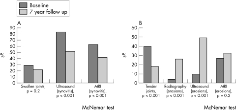
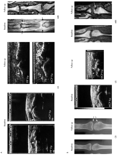
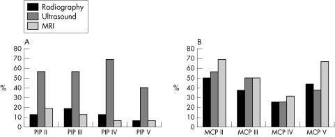
Results: At baseline, CR detected erosions in 5/128 (4%) of all joints, US in 12/128 (9%), and MRI in 34/128 (27%). Seven years later, an increase of joints with erosions was found with CR (26%), US (49%) (p<0.001 each), and MRI (32%, NS). In contrast, joint swelling and tenderness assessed by clinical examination were decreased at follow up (p = 0.2, p<0.001). A significant reduction in synovitis with US and MRI (p<0.001 each) was seen. In CR, 12 patients did not have any erosions at baseline, while in 10/12 patients erosions were detected in 25/96 (26%) joints after 7 years. US initially detected erosions in 9 joints, of which two of these joints with erosions were seen by CR at follow up. MRI initially found 34 erosions, of which 14 (41%) were then detected by CR.
Conclusion: After 7 years, an increase of bone erosions was detected by all imaging modalities. In contrast, clinical improvement and regression of synovitis were seen only with US and MRI. More than one third of erosions previously detected by MRI were seen by CR 7 years later.
Conflict of interest statement
Competing interests: None.
Similar articles
-
Prospective two year follow up study comparing novel and conventional imaging procedures in patients with arthritic finger joints.Ann Rheum Dis. 2002 Oct;61(10):895-904. doi: 10.1136/ard.61.10.895. Ann Rheum Dis. 2002. PMID: 12228160 Free PMC article.
-
Magnetic resonance imaging, radiography, and scintigraphy of the finger joints: one year follow up of patients with early arthritis. The TIRA Group.Ann Rheum Dis. 2000 Jul;59(7):521-8. doi: 10.1136/ard.59.7.521. Ann Rheum Dis. 2000. PMID: 10873961 Free PMC article.
-
Low-cost, low-field dedicated extremity magnetic resonance imaging in early rheumatoid arthritis: a 1-year follow-up study.Ann Rheum Dis. 2006 Sep;65(9):1208-12. doi: 10.1136/ard.2005.049213. Epub 2006 Mar 15. Ann Rheum Dis. 2006. PMID: 16540550 Free PMC article.
-
Imaging in rheumatoid arthritis--why MRI and ultrasonography can no longer be ignored.Scand J Rheumatol. 2003;32(2):63-73. doi: 10.1080/03009740310000058. Scand J Rheumatol. 2003. PMID: 12737323 Review.
-
The clinical features of rheumatoid arthritis.Eur J Radiol. 1998 May;27 Suppl 1:S18-24. doi: 10.1016/s0720-048x(98)00038-2. Eur J Radiol. 1998. PMID: 9652497 Review.
Cited by
-
The utility of MRI in predicting radiographic erosions in the metatarsophalangeal joints of the rheumatoid foot: a prospective longitudinal cohort study.Arthritis Res Ther. 2009;11(3):R94. doi: 10.1186/ar2737. Epub 2009 Jun 22. Arthritis Res Ther. 2009. PMID: 19545417 Free PMC article.
-
Is musculoskeletal ultrasonography an operator-dependent method or a fast and reliably teachable diagnostic tool? Interreader agreements of three ultrasonographers with different training levels.Int J Rheumatol. 2010;2010:164518. doi: 10.1155/2010/164518. Epub 2010 Dec 9. Int J Rheumatol. 2010. PMID: 21197088 Free PMC article.
-
Evaluation of a new erosion score by musculoskeletal ultrasound in patients with rheumatoid arthritis: is US ready for a new erosion score?Clin Rheumatol. 2014 Sep;33(9):1255-62. doi: 10.1007/s10067-014-2646-7. Epub 2014 May 14. Clin Rheumatol. 2014. PMID: 24824913
-
Analysis of distribution and severity of inflammation in patients with osteoarthitis compared to rheumatoid arthritis by ICG-enhanced fluorescence optical imaging and musculoskeletal ultrasound: a pilot study.Ann Rheum Dis. 2016 Mar;75(3):566-70. doi: 10.1136/annrheumdis-2015-207345. Epub 2015 Aug 26. Ann Rheum Dis. 2016. PMID: 26311723 Free PMC article.
-
[New developments in joint ultrasonography].Z Rheumatol. 2006 Dec;65(8):700-7. doi: 10.1007/s00393-006-0120-x. Z Rheumatol. 2006. PMID: 17075711 Review. German.
References
-
- Peterfy C G. Magnetic resonance imaging of rheumatoid arthritis: the evolution of clinical applications through clinical trials. Semin Arthritis Rheum 200130375–396. - PubMed
-
- Bird P, Ejbjerg B, McQueen F, Ostergaard M, Lassere M, Edmonds J. OMERACT Rheumatoid Arthritis Magnetic Resonance Imaging Studies. Exercise 5: an international multicenter reliability study using computerized MRI erosion volume measurements, J Rheumatol 2003301380–1384. - PubMed
Publication types
MeSH terms
LinkOut - more resources
Full Text Sources
Medical
Miscellaneous
