. 2005 Sep 28;25(39):8843-53.
doi: 10.1523/JNEUROSCI.2868-05.2005.

Lipopolysaccharide-induced inflammation exacerbates tau pathology by a cyclin-dependent kinase 5-mediated pathway in a transgenic model of Alzheimer's disease

Affiliations

Lipopolysaccharide-induced inflammation exacerbates tau pathology by a cyclin-dependent kinase 5-mediated pathway in a transgenic model of Alzheimer's disease

Masashi Kitazawa et al. J Neurosci. .

Abstract

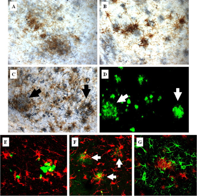
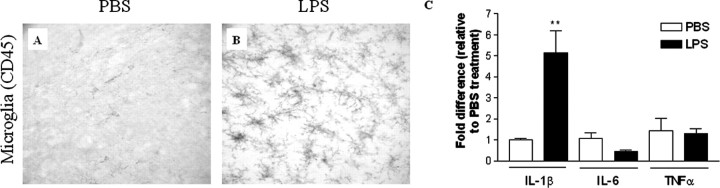
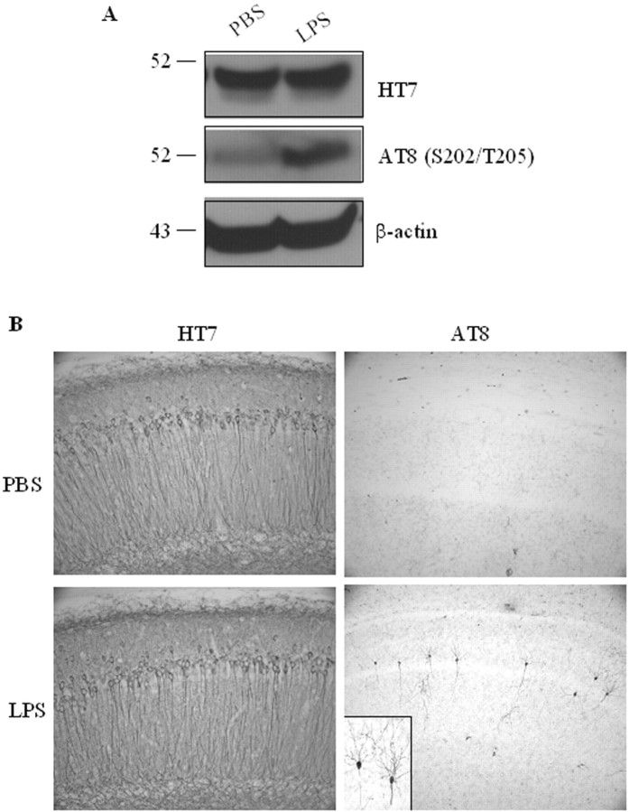
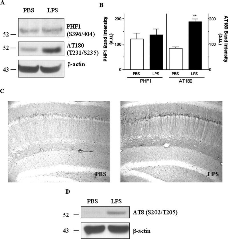
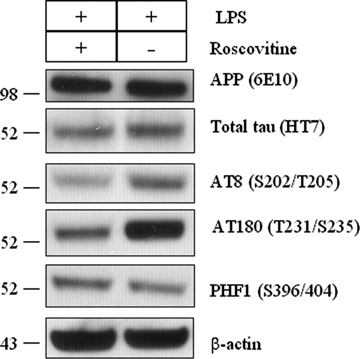
Inflammation is a critical component of the pathogenesis of Alzheimer's disease (AD). Although not an initiator of this disorder, inflammation nonetheless plays a pivotal role as a driving force that can modulate the neuropathology. Here, we characterized the time course of microglia activation in the brains of a transgenic model of AD (3xTg-AD) and discerned its relationship to the plaque and tangle pathology. We find that microglia became activated in a progressive and age-dependent manner, and this activation correlated with the onset of fibrillar amyloidbeta-peptide plaque accumulation and tau hyperphosphorylation. To determine whether microglial activation can exacerbate the pathology, we exposed young 3xTg-AD mice to lipopolysaccharide (LPS), a known inducer of CNS inflammation. Although amyloid precursor protein processing appeared unaffected, we find that LPS significantly induced tau hyperphosphorylation at specific sites that were mediated by the activation of cyclin-dependent kinase 5 (cdk5) through increased formation of the p25 fragment. We further show that administration of roscovitine, a selective and potent inhibitor of cdk5, markedly blocked the LPS-induced tau phosphorylation in the hippocampus. Therefore, this study clearly demonstrates that microglial activation exacerbates key neuropathological features such as tangle formation.


Figures

Figure 1.
Figure 1.
Temporal profile of brain inflammation in the 3xTg-AD mice. The time course of intraneuronal and extraneuronal Aβ buildup and its relationship to activated microglia were described in the 3xTg-AD mice. Both intraneuronal and extraneuronal Aβ deposits were determined by 6E10 immunostaining of 3xTg-AD brains at 9 (A), 12 (B), and 24 (C) months of age. Fibrillar amyloid plaques were detected by thioflavin S staining at 9 (D), 12 (E), and 24 (F) months of age. Serial sections were stained with anti-CD45 to detect activated microglia at 9 (H), 12 (I), and 24 (J) months of age. Amyloid burden (G) and CD45-immunopositive-activated microglia (K) were quantitatively analyzed by measuring the percentage of occupied area by thioflavin S (amyloid burden) or CD45-immunopositive staining (microglia burden). Data are presented as mean±SEM (n=5 in each age). Higher-magnification images of CD45-immunopositive staining in the subicular subfield of the hippocampus from a 24-month-old NonTg mouse (L) and 3xTg-AD mouse (M) are shown. GFAP immunostaining revealed no gliosis in layers III–IV of the neocortex of a 24-month-old NonTg mouse (N), whereas there was a marked increase in GFAP-immunoreactive astrocytes detected in 24-month-old 3xTg-AD mice (O).
Figure 2.
Figure 2.
Activated microglia colocalize with plaques in aged 3xTg-AD mice. Activated microglia detected by CD45 immunoreactivity (brown) colocalize with extraneuronal Aβ-immunopositive deposits (blue) in the cortex (A), amygdala (B), and hippocampus (C) in 24-month-old 3xTg-AD mice. Serial brain sections of the hippocampus were stained with thioflavin S (D). Arrows point to activated microglia around thioflavin S-positive plaques. Additional double-staining fluorescent analysis using thioflavin S (green) and Iba1 (red) to detect microglia demonstrates that 3xTg-AD mice (E) show similar microglia and plaque interactions as observed in the hippocampus of the human AD brain (F). Arrows point to microglia associated with the cores of amyloid plaques in the human AD brain. G, Double fluorescent labeling of GFAP-immunopositive astrocytes (green) and amyloid-containing plaques (red) detected by 6E10 in 24-month-old 3xTg-AD mice shows colocalization of astrocytes around plaques.
Figure 3.
Figure 3.
LPS activates microglia in the 3xTg-AD mice. LPS (0.5 mg/kg body weight) or PBS was injected intraperitoneally 4-month-old 3xTg-AD mice twice per week for 6 weeks, and the brains were analyzed by immunohistochemistry. CD45 immunoreactivity was compared in the hippocampus of PBS- and LPS-treated 3xTg-AD mice. The brains from PBS-treated mice show only basal levels of microglial activation (A), whereas LPS-treated mice show significantly increased activated microglia (B). At least five mice per group were analyzed. C, Quantitative RT-PCR of IL-1β and IL-6 was performed in total RNA extracts from PBS- or LPS-treated 3xTg-AD mice brains. In each mouse, the expression levels of mRNA were normalized relative to the levels of GAPDH mRNA (Ct values for the PBS- and LPS-treated brain are 14.54 ± 0.22 and 14.55 ± 0.18, respectively), and fold differences were calculated relative to the PBS-treated group for each IL. **p < 0.01 compared with the PBS-treated mice group (n = 3 in group).
Figure 4.
Figure 4.
Effect of LPS-induced inflammation on APP processing in the 3xTg-AD mice. A, Immunoblotting of APP, C99, and C83 in PBS- or LPS-treated 3xTg-AD mice, withβ-actin used as a protein loading control. B, Densitometric analysis of steady-state levels of full-length APP, C99, or C83 between PBS- and LPS-treated 3xTg-AD mice. Data are presented as mean ± SEM (n = 4 each group). C, Total Aβ40 and Aβ42 from detergent-soluble and -insoluble fractions of PBS- and LPS-treated 3xTg-AD mice were measured by ELISA. □, PBS-treated 3xTg-AD mice; ▪, LPS-treated 3xTg-AD mice. Data are presented as mean ± SEM (n = 8 each group). D, Fluorescent intensity of 6E10-immunopositive neurons in the cortex and hippocampus was quantitatively analyzed using Quantity One software. Data are presented as mean intensity ± SEM (n = 5 each group). E, Representative immunofluorescent staining of APP and Aβ fragments by 6E10 in the neocortex and hippocampus of 3xTg-AD mice treated with PBS or LPS. a.u., Arbitrary units; HC, hippocampus; CX, cortex.
Figure 5.
Figure 5.
Inflammation-induced enhancement of tau phosphorylation. A, Immunoblotting analysis of total tau (HT7) and phosphorylated tau (AT8) in PBS- and LPS-treated 3xTg-AD mice, with β-actin used as a protein loading control. Data represent at least five mice per group. B, Immunohistochemical staining of total human tau and phosphorylated tau in hippocampal neurons of 3xTg-AD mice treated with PBS and LPS. The inset in the bottom right panel is a magnified view of AT8-immunopositive neurons in the hippocampus.
Figure 6.
Figure 6.
Site-specific phosphorylation of tau after LPS exposure. A, Immunoblotting of AT180- and PHF1-positive phospho-tau in LPS- or PBS-treated 3xTg-AD mice brain, with β-actin used as a protein loading control. B, Densitometric analysis of steady-state levels of PHF1 or AT180 between PBS- and LPS-treated 3xTg-AD mice. Data are presented as mean ± SEM (n = 5). **p < 0.01. C, Representative immunohistochemical staining of AT180-positive tau in hippocampal neurons of 3xTg-AD mice treated with PBS and LPS. D, Six-month-old NonTg mice were treated with 0.5 mg/kg LPS or PBS intraperitoneally twice per week for 6 weeks. After the treatment period, the hippocampus and cortex were isolated, and proteins were analyzed by immunoblotting for AT8-positive phosphorylated tau. a.u., Arbitrary units.
Figure 7.
Figure 7.
LPS-induced inflammation causes increased p25 formation. A, Immunoblotting for p35/p25, cdk5, phosphorylated/total GSK-3β, phosphorylated/total p38–MAPK, and phosphorylated/total JNK of PBS- and LPS-treated 3xTg-AD mice, withβ-actin used as a protein loading control. Densitometric analysis revealed a significant (p < 0.05) increase in p25 levels in LPS-treated 3xTg-AD mice (n = 5; data not shown). B, Immunoblotting for p35/p25 and cdk5 of PBS- and LPS-treated NonTg mice, with β-actin used as a protein loading control. Densitometric analysis revealed a significant (p < 0.05) increase in p25 levels in LPS-treated NonTg mice (n = 4; data not shown).
Figure 8.
Figure 8.
Roscovitine inhibits the cdk5-dependent phosphorylation of tau in LPS-treated 3xTg-AD mice. 3xTg-AD mice were treated with roscovitine (300 nmol per day) or vehicle (50% DMSO) through an intraventricular infusion during the last 2 weeks of the 6 week LPS treatment. The hippocampus was isolated and tested for phosphorylation status of tau by immunoblotting. The steady-state levels of human APP and tau were not altered by roscovitine treatment. Densitometric analysis revealed the steady-state levels of AT8- and AT180-positive tau resulted in nearly a twofold and threefold reduction in roscovitine treatment, respectively, whereas PHF1-positive tau levels were not affected by LPS (data not shown). Membranes were reprobed with β-actin to control the equal protein loading (n = 3 each group).

References

    1. Ahlijanian MK, Barrezueta NX, Williams RD, Jakowski A, Kowsz KP, McCarthy S, Coskran T, Carlo A, Seymour PA, Burkhardt JE, Nelson RB, McNeish JD (2000) Hyperphosphorylated tau and neurofilament and cytoskeletal disruptions in mice overexpressing human p25, an activator of cdk5. Proc Natl Acad Sci USA 97: 2910–2915. - PMC - PubMed
    1. Akiyama H, Barger S, Barnum S, Bradt B, Bauer J, Cole GM, Cooper NR, Eikelenboom P, Emmerling M, Fiebich BL, Finch CE, Frautschy S, Griffin WS, Hampel H, Hull M, Landreth G, Lue L, Mrak R, Mackenzie IR, McGeer PL, et al. (2000) Inflammation and Alzheimer's disease. Neurobiol Aging 21: 383–421. - PMC - PubMed
    1. Alvarez A, Toro R, Caceres A, Maccioni RB (1999) Inhibition of tau phosphorylating protein kinase cdk5 prevents beta-amyloid-induced neuronal death. FEBS Lett 459: 421–426. - PubMed
    1. Anthony JC, Breitner JC, Zandi PP, Meyer MR, Jurasova I, Norton MC, Stone SV (2000) Reduced prevalence of AD in users of NSAIDs and H2 receptor antagonists: the Cache County study. Neurology 54: 2066–2071. - PubMed
    1. Apelt J, Schliebs R (2001) Beta-amyloid-induced glial expression of both pro- and anti-inflammatory cytokines in cerebral cortex of aged transgenic Tg2576 mice with Alzheimer plaque pathology. Brain Res 894: 21–30. - PubMed

Publication types

MeSH terms