. 2005 Sep 28;25(39):8878-88.
doi: 10.1523/JNEUROSCI.2005-05.2005.

Calcium/calmodulin-dependent protein kinase II alters structural plasticity and cytoskeletal dynamics in Drosophila

Affiliations

Calcium/calmodulin-dependent protein kinase II alters structural plasticity and cytoskeletal dynamics in Drosophila

Ryan Andersen et al. J Neurosci. .

Abstract

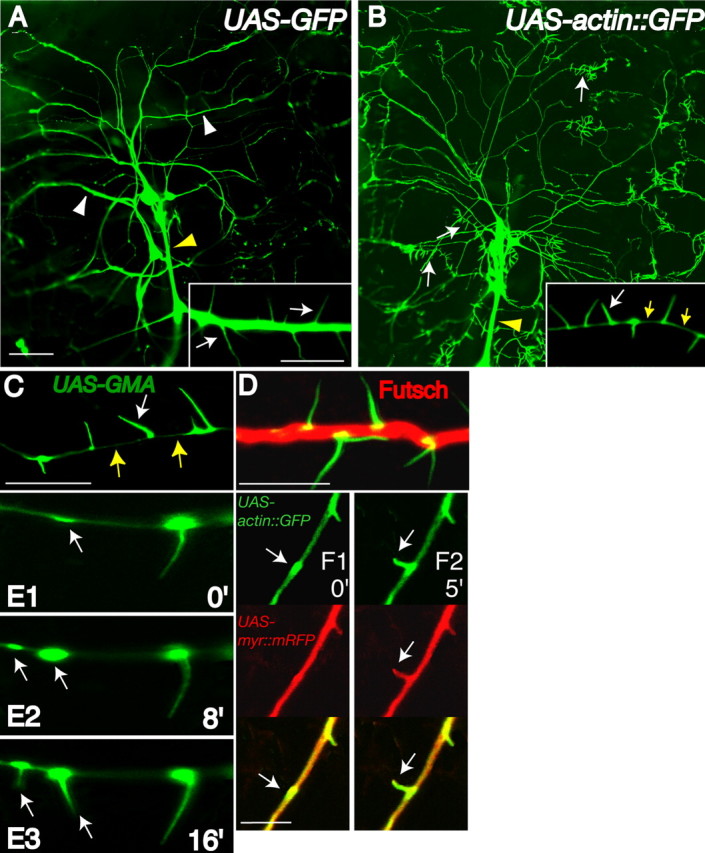
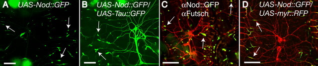
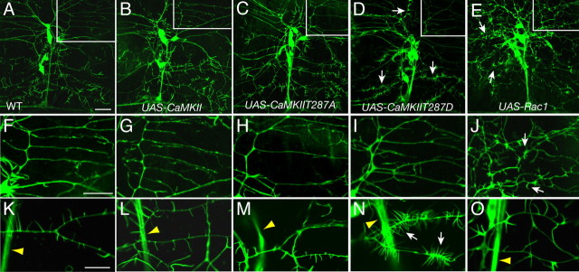
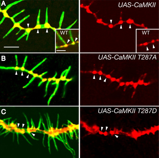
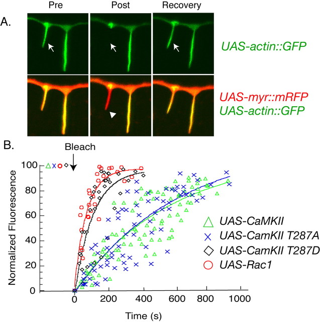
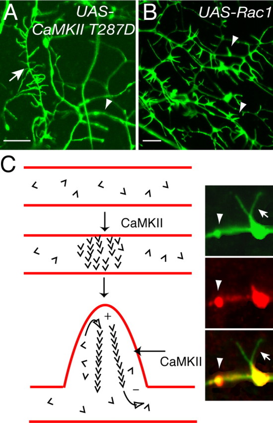
Drosophila dendritic arborization (da) neurons contain subclasses of neurons with distinct dendritic morphologies. We investigated calcium/calmodulin-dependent protein kinase II (CaMKII) regulation of dendritic structure and dynamics in vivo using optically transparent Drosophila larvae. CaMKII increases the dynamic nature and formation of dendritic filopodia throughout larval development but only affects neurons that normally contain dendritic filopodia. In parallel, we examined the effects of Rac1 activity on dendritic structure to explore signaling specificity. In contrast to CaMKII activity, Rac1 does not alter filopodia stability but instead causes de novo filopodia formation on all da neurons. Although both mediators increase cytoskeletal turnover, measured by fluorescence recovery after photobleaching experiments, only CaMKII increases the dynamic nature of dendritic filopodia. CaMKII signaling thus appears to use mechanisms and machinery distinct from Rac1 signaling. This study illustrates a molecular means of uncoupling cytoskeletal regulation from morphological regulation. Our results suggest that Drosophila dendritic filopodia may share some cytoskeletal regulatory mechanisms with mammalian dendritic filopodia. Furthermore, general dendrite cytoskeletal compartmentalization is conserved in multipolar neurons.


Figures

Figure 1.
Figure 1.
Drosophila da neurons contain actin-rich filopodia restricted to dendrite compartments. Single-neuron dendrite images (A–F) are from the ddaA neuron from the dorsal cluster of sensory neurons. All images throughout this study contain hemi-segment A6 dorsal cluster da neurons with anterior toward the left and dorsal toward the top. A, A second instar larva (yw; Gal4–109(2)80, UAS-GFP) expressing GFP in da neurons demonstrates strong dendritic shaft (white arrowheads) and axon fascicle (yellow arrowhead) labeling. B, In comparison, a second instar larva (yw; Gal4–109(2)80, UAS-actin::GFP) expressing actin::GFP reveals actin-rich dendritic filopodia along dendrites (white arrows) that are absent on axonal shafts (yellow arrowhead). Actin::GFP demonstrates strong enrichment in dendritic filopodia with only limited fluorescence in dendritic shafts (inset, yellow arrows). C, When a second instar larva (yw; Gal4–109(2)80, UAS-GMA) expresses GMA, an F-actin-binding GFP fusion protein, dendritic filopodia are strongly labeled with faint localization in dendritic shafts (yellow arrows). D, A transgenic second instar larva expressing GMA immunolabeled against Futsch (red), a microtubule-binding protein, GMA (α-GFP; green), reveals mostly separate dendritic localization for microtubule- and actin-associated proteins. E1–E3, In vivo 8 min time-lapse images of a GMA expressing transgenic second instar larva indicate early recruitment of F-actin to dendritic patches that predict sites of future dendritic filopodia outgrowth (arrows) (time in minutes). F1, F2, In vivo 5 min time-lapse images of a second instar larva (yw; Gal4–109(2)80, UAS-actin::GFP, UAS-myr::mRFP) expressing actin:GFP (green; arrows) and myr::mRFP reveal membrane outgrowth (red; arrows) is closely associated with actin::GFP outgrowth (green; arrows). Scale bars: (in A) A, B, 50 μm; insets, 5 μm; C, D, F, 5 μm.
Figure 2.
Figure 2.
Asynaptic Drosophila da neurons contain minus-end-out microtubule arrays in dendrites. A, A second instar larva (yw; Gal4 109(2)80, UAS-Nod:GFP) expressing Nod:GFP, a minus-end reporter for microtubules, demonstrates Nod:GFP enrichment at the tips of da dendrites (arrows). B, A second instar larva (yw; Gal4–109(2)80, UAS-Nod::GFP/Gal4–109(2)80 UAS-Tau::GFP) heterozygous for both Tau::GFP, a microtubule-binding protein that labels dendrites, and Nod::GFP demonstrates that Nod::GFP accumulates at the ends of dendrites collinear with the dendrite shaft. C, A second instar larva (yw; Gal4 109(2)80, UAS-Nod::GFP) immunolabeled against Nod::GFP (αGFP; green) and Futsch (αFutsch; red) reveals colabeling (yellow) as puncta at the ends of da neuronal dendrites (arrows). D, A second instar larva (yw; Gal4 109(2)80 UAS-myr::RFP/UAS-Nod::GFP) heterozygous for both myr:RFP and Nod::GFP demonstrates Nod:GFP accumulation at the ends of da dendrites (arrows). Scale bars, 50 μm.
Figure 3.
Figure 3.
CaMKII T287D expression in transgenic larvae induces high densities of filopodia on class III neuronal dendrites, whereas Rac1 expression induces filopodia formation on non-class III da neuronal dendrites. All larvae are second instars, homozygous for Gal4–109(2)80, UAS-actin::GFP and UAS-CaMKII variants or UAS-Rac1 transgenes. A, F, K, Dendrites from a wild-type (WT) larva are indistinguishable from a transgenic larva expressing wild-type CaMKII (B, G, L) and nearly identical to a larva expressing CaMKII T287A (C, H, M). D, I, N, In contrast, a transgenic larva expressing CaMKII T287D demonstrates striking increases in numbers of filopodia on class III da neuronal dendrites (arrows). E, J, O, Rac1 expression in a transgenic larva causes increased dendritic branching and filopodia formation on all da neurons. F–J, Boxed regions (above, A–E) reveal that a class I da neuron, ddaE, does not have filopodia when expressing any protein variant except Rac1 (J, arrows). K–O, Equivalent dendritic branches from the class III ddaA neuron have marked increases in dendritic filopodia when expressing CaMKII T287D (N) compared with WT (K), CaMKII (L), CaMKII T287A (M), or Rac1 (O). Note that axon shaft fascicles (yellow arrowheads) in all genotypes lack filopodia. Scale bars: (in A, F) A–F, 20 μm; (in F) F–J, 10 μm; (in K) K–O, 10 μm. All larvae for the remaining figure legends are of the background genotype yw; Gal4 109(2)80, UAS-actin:GFP to allow visualization of dendritic filopodia.
Figure 4.
Figure 4.
Class III dendrites of transgenic larvae expressing CaMKII T287D contain increasing filopodia in a gene-copy-dependent manner, whereas CaMKII T287D activity, but not Rac1 activity, modulates dendritic filopodia stability. A, Second instar larvae expressing CaMKII T287D possess increasing numbers of filopodia on ddaA neurons with increasing transgene number. Expression of CaMKII T287A shifts the distribution toward slightly longer filopodia (#p < 0.0001; χ2 test; n = 7 of wild-type CaMKII and CaMKII T287A) but does not increase total number (*p < 0.06; **p < 0.006; ***p < 0.0007; t test; n = 7 of each genotype; all filopodia from the entire ddaA dendritic arbor were counted.) B, Time-lapse imaging (2–3 min intervals; total, 15 min) of the ventral dendrite segment (100μm) of neuron ddaA demonstrates that CaMKII T287D, but not Rac1, activity increases formation and disappearance of filopodia (*p < 0.02; ***p < 8 × 10⁻⁶). C, CaMKII expression decreases the stable pool of filopodia when compared with WT, WT CaMKII, CaMKII T287A, and Rac1 [**p < 0.004; n = 7; 25 filopodia from a 100μm ventral dendrite segment of neuron ddaA segment (similar toB) were imaged every2–3min for 15 min. The filopodia were then classified as stable, elongating/retracting, or disappeared. Seven animals for each genotype were quantified]. Error bars represent SEM. D–F, Newly hatched first instar larvae and third instar larvae (G–I) reveal timing differences between CaMKII T287D and Rac1 mutant phenotypes. WTCaMKII (D) and CaMKII T287D (E) first instar larvae appear similar. In contrast, a Rac1 (F)-expressing larva shows an early branching and filopodia mutant phenotype. G–I, Third instar larvae class III neuron ddaF demonstrates CaMKII T287D expression has a cumulative phenotype (H), consistent with continuous increased filopodia formation (B). The Rac1 phenotype appears relatively stable, with increased branching most prominent. Scale bars: D, G,10 μm. WT, Wild type.
Figure 5.
Figure 5.
CaMKII protein variants show enrichment at the intersection of F-actin and microtubule compartments with similar dendritic subcellular localization in fixed specimens. A–C, Anti-CaMKII immunolabeling (red) of endogenous CaMKII (A, inset) expressed wild-type (WT) CaMKII (A), CaMKII T287A (B), or CaMKII T287D (C) in second instar larvae of neuron ddaA. CaMKII enrichment (arrowheads) is found at the intersection of dendrites and filopodia (anti-GFP; green projections). Note the phenotype in CaMKII T287D (C) and the association of individual filopodia with CaMKII-immunopositive clusters in all larvae. Scale bars: A,5 μm.
Figure 6.
Figure 6.
Intact CaMKII T287D and wild-type Rac1 transgenic larvae demonstrate significantly increased actin::GFP turnover in dendritic filopodia. A, Actin::GFP turnover in dendritic filopodia was demonstrated using fluorescence recovery after FRAP (arrows). A transgenic animal containing both UAS-actin::GFP and membrane-targeted UAS-myr::mRFP reveals that FRAP itself does not visibly affect visible filopodia structure (arrowhead). Pre, Prediffusion; Post, postdiffusion. B, Individual data points from seven animals of each genotype were fit to F (t) = 1 – fsffet, an equation used previously to document FRAP for actin::GFP in dendritic spines of transfected hippocampal neurons (Star et al., 2002). Actin::GFP turnover in UAS-CaMKII T287D (t1/2 = 132 s; p < 2 × 10⁻⁶; black diamond) and UAS-Rac1 (t1/2 102 s; p < 6×10⁻⁶; red circle) animals dramatically increased compared with UAS-CaMKII T287A (t1/2 = 347 s; blue cross) or UAS-CaMKII animals (green triangles).
Figure 7.
Figure 7.
Rac1 and CaMKII modulate dendritic filopodia by distinct mechanisms. A, B, Analyzing dendrites of class I neuron ddaE (A, arrowhead) and class III neuron ddaF (A, arrow) demonstrates that CaMKII only affects neurons that normally contain filopodia (A, arrow), leaving neighboring dendrites from nonfilopodia-containing neurons (A, arrowhead) unaffected. In contrast, Rac1 activity (B) causes de novo dendritic filopodia formation on all dendritic arborization neurons regardless of class types (B, arrowheads). Scale bars, 5 μm. C, Schematic model of dendritic effects mediated by activated CaMKII on dendritic filopodia and actin turnover. A patch of dendritic membrane uniquely accumulates small amounts of filamentous actin. This patch can be the site of future filopodia extension (Fig. 1 E). Free actin monomers (barbs) are continuously added to the filaments at the plus (+) end and can become free monomer by dissociation at the minus (–) end. CaMKII could increase the exchange rate of monomers to result in increased rates of fluorescence recovery after photobleaching and also increase the formation of actin-membrane patches. Inset, Colocalization of CaMKII (red) and actin:GFP (green) in dendrites. CaMKII accumulates in dendrites specifically at sites of F-actin enrichment and dendritic filopodia formation.

References

    1. Ainsley JA, Pettus JM, Bosenko D, Gerstein CE, Zinkevich N, Anderson MG, Adams CM, Welsh MJ, Johnson WA (2003) Enhanced locomotion caused by loss of the Drosophila DEG/ENaC protein Pickpocket1. Curr Biol 13: 1557–1563. - PubMed
    1. Baas PW, Deitch JS, Black MM, Banker GA (1988) Polarity orientation of microtubules in hippocampal neurons: uniformity in the axon and nonuniformity in the dendrite. Proc Natl Acad Sci USA 85: 8335–8339. - PMC - PubMed
    1. Bodmer R, Jan YN (1987) Morphological differentiation of the embryonic peripheral neurons in Drosophila. Rouxs Arch Dev Biol 196: 69–77. - PubMed
    1. Brand AH (1995) GFP in Drosophila. Trends Genet 11: 324–325. - PubMed
    1. Brand AH, Perrimon N (1993) Targeted gene expression as a means of altering cell fates and generating dominant phenotypes. Development 118: 401–415. - PubMed

Publication types

MeSH terms

Substances

LinkOut - more resources