. 2005 Sep 28;25(39):8978-87.
doi: 10.1523/JNEUROSCI.2246-05.2005.

Hippocampal inactivation disrupts the acquisition and contextual encoding of fear extinction

Affiliations

Hippocampal inactivation disrupts the acquisition and contextual encoding of fear extinction

Kevin A Corcoran et al. J Neurosci. .

Erratum in

  • J Neurosci. 2005 Oct 19;25(42):9821

Abstract

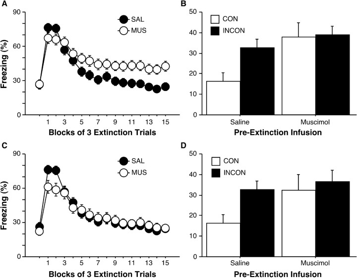
In recent studies, inactivation of the dorsal hippocampus before the retrieval of extinguished fear memories disrupted the context-dependent expression of these memories. In the present experiments, we examined the role of the dorsal hippocampus in the acquisition of extinction. After pairing an auditory conditional stimulus (CS) with an aversive footshock [unconditional stimulus (US)], rats received an extinction session in which the CS was presented without the US. In experiment 1, infusion of muscimol, a GABAA receptor agonist, into the dorsal hippocampus before the extinction training session decreased the rate of extinction. Moreover, when later tested for fear to the extinguished CS, all rats that had received hippocampal inactivation before extinction training demonstrated renewed fear regardless of the context in which testing took place. This suggests a role for the dorsal hippocampus in both acquiring the extinction memory and encoding the CS-context relationship that yields the context dependence of extinction. In experiment 2, inactivation of the dorsal hippocampus before testing also disrupted the context dependence of fear to the extinguished CS. In experiment 3, quantitative autoradiography revealed the boundaries of muscimol diffusion after infusion into the dorsal hippocampus. Together, these results reveal that the dorsal hippocampus is involved in the acquisition, contextual encoding, and context-dependent retrieval of fear extinction. Learning and remembering when and where aversive events occur is essential for adaptive emotional regulation.


Figures

Figure 1.
Figure 1.
Cannula placement in the dorsal hippocampus (experiment 1). Photomicrograph showing a thionin-stained coronal section from the brain of a rat with representative cannula placements in the dorsal hippocampus.
Figure 2.
Figure 2.
Illustration of guide cannula placements in the dorsal hippocampus (experiment 1). Placements represented are from most rats included in the final analysis; many rats were transported to another laboratory for use in another experiment (INCON/SAL, filled squares; INCON/MUS, open squares; CON/SAL, filled circles; CON/MUS, open circles). Atlas templates were adapted from Swanson (1992).
Figure 3.
Figure 3.
Hippocampal inactivation disrupts the acquisition and contextual encoding of extinction (experiment 1). A, Freezing across the 3 pre-CS minutes and 45 post-CS minutes during the extinction training session. Rats were given infusions of MUS (open circles) or SAL (filled circles) before extinction training. B, Freezing across the first 6 post-CS minutes of the testing session. Rats were tested in a context that was either consistent (open bars) or inconsistent (filled bars) with extinction training. Muscimol and saline refer to infusions that were given before extinction training. C, Freezing during the extinction training session among rats matched for levels of freezing at the end of the extinction training session. D, Freezing during the testing session for rats matched for levels of freezing at the end of the extinction training session. All data are presented as means ± SEM.
Figure 4.
Figure 4.
Effects of hippocampal inactivation on extinction training and testing did not differ across contexts. A, Freezing behavior averaged across the first 3, middle 3, and final 3 post-CS minutes of the extinction training session. All rats were fear conditioned in context A. Extinction training occurred the next day, in either the same context as conditioning (A→A; circles) or a new context (context B; A→B; squares) after infusions of either muscimol (open symbols) or saline (filled symbols). B, Renewal of fear to the CS was similar among all INCON/SAL, CON/MUS, and INCON/MUS groups, relative to the CON/SAL group (CON groups, open bars; INCON groups, filled bars). Muscimol and saline refer to infusions given before extinction training. All data are presented as means ± SEM.
Figure 5.
Figure 5.
Hippocampal inactivation before testing disrupts renewal of fear after a single extinction training session (experiment 2). Mean ± SEM percentage of freezing averaged across the first 5 post-CS minutes during testing. Rats were tested either in a context consistent with extinction training (open bars) or in a context inconsistent with CS-alone presentations during the extinction training (filled bars). Testing took place 20–25 min after an intrahippocampal infusion of either saline or muscimol.
Figure 6.
Figure 6.
Location of [3H]muscimol at various intervals after intrahippocampal infusions (experiment 3). Phosphoimages of [3H]muscimol were made and overlaid on postfixed cresyl-violet-stained sections. Rats were killed immediately (0 min), 20, 60, or 360 min after intrahippocampal [3H]muscimol infusions. Sections shown are from representative rats from each infusion–killing interval and range from 0.6 mm rostral (leftmost section) to 0.6 mm caudal (rightmost section) to the infusion site.
Figure 7.
Figure 7.
Diffusion of [3H]muscimol away from the site of infusion (experiment 3). Mean ± SEM apparent density from tissue sections taken throughout the rostrocaudal extent of the hippocampus. Sections from each rat having the maximum density throughout the infused area were placed at distance zero; all other sections were placed according to their relative distances from those sections. Rats were killed at one of four time intervals after [3H] muscimol infusion: immediately (0 min; white circles), 20 min (light gray circles), 60 min (dark gray circles), or 360 min (black circles).
Figure 8.
Figure 8.
Spread of [3H]muscimol in the hippocampus (experiment 3). A, Extent of rostrocaudal spread of [3H]muscimol through the hippocampus. Mean ± SEM distance from the first to the last of the tissue sections taken in which radioactive signal was apparent. B, Mean ± SEM two-dimensional area of [3H]muscimol spread in the tissue section having the maximum apparent radioactive density. This section was assumed to be at or near the center of the infusion. Rats were killed at one of four time intervals after [3H]muscimol infusion: immediately (0 min; white bars), 20 min (light gray bars), 60 min (dark gray bars), or 360 min (black bars).

References

    1. Benoit SC, Davidson TL, Chan K-H, Trigilio T, Jarrard LE (1999) Pavlovian conditioning and extinction of context cues and punctate CSs in rats with ibotenate lesions of the hippocampus. Psychobiology (Austin, Tex) 27: 26–39.
    1. Bouton ME (1993) Context, time, and memory retrieval in the interference paradigms of pavlovian conditioning. Psychol Bull 114: 80–99. - PubMed
    1. Bouton ME (1994) Context, ambiguity, and classical conditioning. Curr Dir Psychol Sci 3: 49–53.
    1. Bouton ME, Bolles RC (1979) Contextual control of the extinction of conditioned fear. Learn Motiv 10: 445–466.
    1. Bouton ME, King DA (1983) Contextual control of the extinction of conditioned fear: tests for the associative value of the context. J Exp Psychol Anim Behav Process 9: 248–265. - PubMed

Publication types

MeSH terms

LinkOut - more resources