. 2005 Sep 28;33(17):5622-32.
doi: 10.1093/nar/gki869. Print 2005.

Characterization of the bisintercalative DNA binding mode of a bifunctional platinum-acridine agent

Affiliations

Characterization of the bisintercalative DNA binding mode of a bifunctional platinum-acridine agent

Jayati Roy Choudhury et al. Nucleic Acids Res. .

Abstract

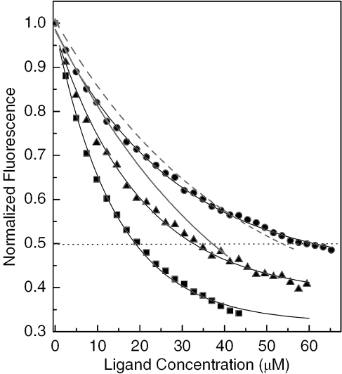
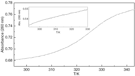
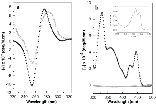
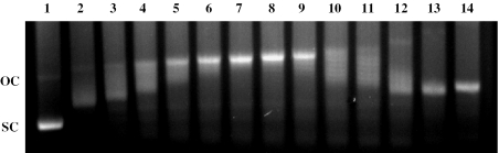
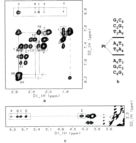
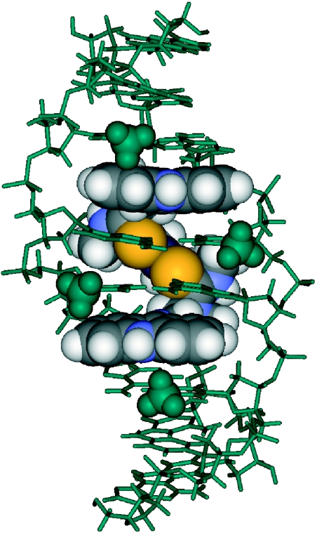
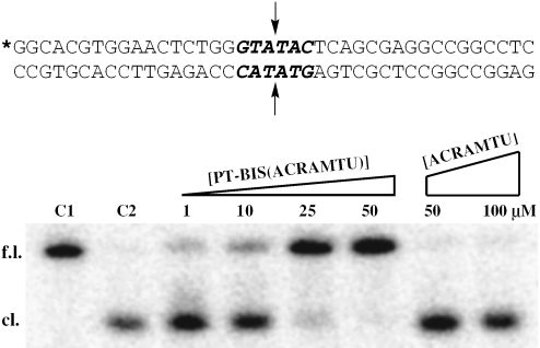
The DNA interactions of PT-BIS(ACRAMTU) ([Pt(en)(ACRAMTU)2](NO3)4; ACRAMTU = 1-[2-(acridin-9-ylamino)ethyl]-1,3-dimethylthiourea, en = ethylenediamine), a bifunctional platinum-acridine conjugate, have been studied in native and synthetic double-stranded DNAs and model duplexes using various biophysical techniques. These include ethidium-DNA fluorescence quenching and thermal melting experiments, circular dichroism (CD) spectroscopy and plasmid unwinding assays. In addition, the binding mode was studied in a short octamer by NMR spectroscopy in conjunction with molecular modeling. In alternating copolymers, PT-BIS(ACRAMTU) shows a distinct preference for poly(dA-dT)2, which is approximately 3-fold higher than that of ACRAMTU. In the ligand-oligomer complex, d(GCTATAGC)2.PT-BIS(ACRAMTU) (complex I*), PT-BIS(ACRAMTU) increases the thermal stability of the B-form host duplex by DeltaT(m) > 30 K (CD and UV melting experiments). The agent unwinds pSP73 plasmid DNA by 44(+/-2) degrees per bound molecule, indicating bisintercalative binding. A 2-D NMR study unequivocally demonstrates that PT-BIS(ACRAMTU)'s chromophores deeply bisintercalate into the 5'-TA/TA base pair steps in I*, while the platinum linker lies in the minor groove. An AMBER model reflecting the NMR results shows that bracketing of the central AT base pairs in a classical nearest neighbor excluded fashion is feasible. PT-BIS(ACRAMTU) inhibits DNA hydrolysis by BstZ17 I at the enzyme's restriction site, GTA downward arrowTAC. Possible consequences for other relevant DNA-protein interactions, such as those involved in TATA-box-mediated transcription initiation and the utility of the platinum-intercalator technology for the design of sequence-specific agents are discussed.


Figures

Figure 1
Figure 1
Structures of PT-BIS(ACRAMTU) and related complexes and sequence I giving atom and residue numberings.
Figure 2
Figure 2
DNA-ethidium fluorescence monitored at 603 nm in the presence of varying concentrations of intercalating agents (numbers in parentheses are C50 [µM] values). Squares: PT-BIS(ACRAMTU)/poly(dA-dT)2 (17.72); triangles: PT-BIS(ACRAMTU)/calf thymus DNA (32.65); circles: PT-BIS(ACRAMTU)/poly(dG-dC)2 (65.14); solid line: ACRAMTU/poly(dA-dT)2 (40.54); dashed line: ACRAMTU/poly(dG-dC)2 (53.57). For clarity, only the fitted curves are shown for ACRAMTU.
Figure 3
Figure 3
UV melting profile monitored at 260 nm for complex I*. The inset shows the melting trace recorded for the unmodified sequence, I. Experiments were performed at octamer concentrations of 3.5 and 7.0 µM, respectively [10 mM phosphate and 10 mM NaCl (pH 7.0)].
Figure 4
Figure 4
CD spectra recorded at 298 K of the modified and unmodified octamer. (a) CD in the DNA region for I (open circles) and I* (filled circles). (b) ICD in the ligand region of I* (filled triangles). The trace close to baseline was recorded for a buffered solution of drug in the absence of oligomer. The inset shows the UV-visible feature of the acridine chromophores in PT-BIS(ACRAMTU). Concentrations of oligomers were 6.3 and 100 µM in (a) and (b), respectively [10 mM phosphate and 10 mM NaCl (pH 7.0)].
Figure 5
Figure 5
Unwinding of supercoiled pSP73 plasmid by PT-BIS(ACRAMTU). With increasing concentration of PT-BIS(ACRAMTU), relaxation of negatively supercoiled plasmid (sc) occurs resulting in the open-circular form (oc), as indicated by the change in electrophoretic mobility of the plasmid. At higher concentrations, PT-BIS(ACRAMTU) induces positive supercoils, thereby increasing the mobility of the modified plasmid. Drug-to-nucleotide ratios (rb) for lanes 1–14 are 0 (control), 0.011, 0.014, 0.017, 0.019, 0.022, 0.025, 0.028, 0.031, 0.034, 0.037, 0.045, 0.056 and 0.068 respectively.
Figure 6
Figure 6
2-D NOESY study of the drug–octamer complex I* [500 MHz, 328 K, τm = 350 ms, buffer: D2O, 10 mM phosphate (pH* 7.0) and 90 mM NaCl]. (a) Section of the 2-D NOESY spectrum showing sequential connectivities in the aromatic-H2′/H2″ region (solid lines). Absent cross-peaks due to disruption of the T3H2′-A4H8 and T5H2′-A6H8 interresidue NOEs are indicated by arrows. The dashed vertical line indicates the connectivity between A4H8 and T5CH3 at the central base pair step. Assignment of intermolecular cross-peaks: (A) A4,A6H2′/H2″-ACRH2/H7; (B) T3H2″-ACRH7; (C) T5H2″-ACRH2; (D) T3H2′-ACRH7; (E) T3CH3-ACRH7; (F) T3H2′-ACRH6; (G) A6H2′/H2″-ACRH1; (H) T5H2″-ACRH1; (I) T3CH3-ACRH5 and T3CH3-ACRH6. The cross-peak for T5CH3- ACRH5 is partially overlapped with the intranucleobase cross-peak, T5CH3-T5H6 (top of the two cross-peaks connected by dashed line). (b) Illustration of the intercalation mode leading to interruption of the NOE connectivities at the 5′-T3A4/T5A6 base pair steps. (c) Section of the 2-D NOESY spectrum showing cross-peaks due to NOE contacts between the thiourea methyl groups (C11) and H1′ and H4′ deoxyribose protons in the minor groove of the octamer. Cross-peak assignments: (A) A4H1′-ACRH11; (B) A6H1′-ACRH11; (C) T3H1′-ACRH11; (D) T5H1′-ACRH11; (E) A6H4′- ACRH11.
Figure 7
Figure 7
Summary of crucial intermolecular and interresidue NOEs observed at the drug binding site in complex I*.
Figure 8
Figure 8
View into the major groove of an energy-minimized AMBER model of duplex I* based on NMR data. The oligomer is shown in green, and the drug molecule is represented by CPK rendering. The thymine methyl groups are highlighted as space-filling spheres.
Figure 9
Figure 9
Restriction enzyme-mediated cleavage by BstZ17 I of a 40 bp duplex modified with PT-BIS(ACRAMTU) or ACRAMTU monitored by denaturing PAGE. Control lanes labeled C1 and C2 contain unmodified probe with no enzyme added and unmodified probe cleaved with enzyme, respectively. Bands are labeled ‘f.l.’ for full-length and ‘cl.’ for cleaved probe. The sequence of the probe is shown with the blunt-end restriction site highlighted in bold. The asterisk denotes the 32P-labeled 5′ end, and the arrows indicate the sites of hydrolytic cleavage.

Similar articles

Cited by

References

    1. Chaires J.B. Drug–DNA interactions. Curr. Opin. Struct. Biol. 1998;8:314–320. - PubMed
    1. Gago F. Stacking interactions and intercalative DNA binding. Methods. 1998;14:277–292. - PubMed
    1. Le Pecq J.B., Le Bret M., Barbet J., Roques B. DNA polyintercalating drugs: DNA binding of diacridine derivatives. Proc. Natl Acad. Sci. USA. 1975;72:2915–2919. - PMC - PubMed
    1. Foster B.J., Clagett-Carr K., Shoemaker D.D., Suffness M., Plowman J., Trissel L.A., Grieshaber C.K., Leyland-Jones B. Echinomycin: the first bifunctional intercalating agent in clinical trials. Invest. New Drugs. 1985;3:403–410. - PubMed
    1. Leroy J.L., Gao X.L., Misra V., Gueron M., Patel D.J. Proton exchange in DNA-luzopeptin and DNA-echinomycin bisintercalation complexes: rates and processes of base-pair opening. Biochemistry. 1992;31:1407–1415. - PubMed

Publication types

Substances