Comparative Study
. 2005 Oct;167(4):1119-24.
doi: 10.1016/S0002-9440(10)61200-X.

Trypsin-2 degrades human type II collagen and is expressed and activated in mesenchymally transformed rheumatoid arthritis synovitis tissue

Affiliations
Comparative Study

Trypsin-2 degrades human type II collagen and is expressed and activated in mesenchymally transformed rheumatoid arthritis synovitis tissue

Mathias Stenman et al. Am J Pathol. 2005 Oct.

Abstract

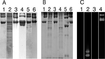
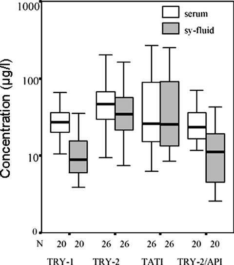
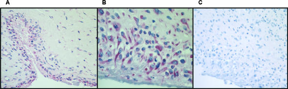
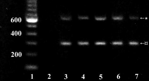
It has traditionally been believed that only the human collagenases (matrix metalloproteinase-1, -8, and -13) are capable of initiating the degradation of collagens. Here, we show that human trypsin-2 is also capable of cleaving the triple helix of human cartilage collagen type II. We purified human trypsin-2 and tumor-associated trypsin inhibitor by affinity chromatography whereas collagen type II was purified from cartilage extracts using pepsin digestion and salt precipitation. Degradation of type II collagen and gelatin by trypsin-2 was demonstrated with sodium dodecyl sulfate-polyacrylamide gel electrophoresis, zymography, and mass spectrometry, and tumor-associated trypsin inhibitor specifically inhibited this degradation. Although human trypsin-2 efficiently digested type II collagen, bovine trypsin did not. Furthermore, immunohistochemical staining detected trypsin-2 in the fibroblast-like synovial lining and in stromal cells of human rheumatoid arthritis synovial membrane. These findings were confirmed by reverse transcriptase-polymerase chain reaction and nucleotide sequencing. Trypsin-2 alone and complexed with alpha(1)-proteinase inhibitor were also detected in the synovial fluid of affected joints by time-resolved immunofluorometric assay, suggesting that trypsin-2 is activated locally. These results are the first to assess the ability of human trypsin to cleave human type II collagen. Thus, trypsin-2 and its regulators should be further studied for use as markers of prognosis and disease activity in rheumatoid arthritis.


Figures

Figure 1
Figure 1
A: Sodium dodecyl sulfate-polyacrylamide gel electrophoresis demonstrates that TRY-2 is an interstitial collagenase able to cleave type II collagen at multiple sites to small molecular weight fragments. Lane 1: Human homotrimeric collagen II substrate only, intact α2 chains. Lane 2: TRY-2 cleaves intact, triple helical human type II collagen monomers at multiple sites to small molecular weight collagen fragments. Lane 3: TRY-2-mediated collagenolysis inhibited by the presence of its specific inhibitor TATI. Lane 4: MMP-8 (neutrophil collagenase, collagenase-2), a positive collagenase control, produces classical ¾ and ¼ fragments as a result of action on the single initial cleavage site for mammalian collagenases. Lanes 5 and 6: MMP-9 (gelatinase B, 92-kd type IV collagenase; lane 5) and bovine trypsin (lane 6) not able to cleave type II collagen. B: Lane 1: Intact type II collagen. Lane 2: TRY-2 cleaves type II collagen. Lane 3: TRY-2 collagenolysis inhibited by TATI. Lane 4: MMP-inhibitor GM 6001 (Ilomastat) does not inhibit TRY-2-mediated collagenolysis of type II collagen. Lanes 5 and 6: MMP-8 produces classic ¾ and ¼ fragments of type II collagen (lane 5) that is inhibited by GM 6001 (Ilomastat) (lane 6). C: Lane 1: Zymography gels demonstrate gelatin substrate only. Lane 2: TRY-2 is also a gelatinase able to cleave gelatin (denatured collagen). Lanes 3 and 4: MMP-8 is not gelatinolytic (lane 3), whereas MMP-9 is gelatinolytic (lane 4). It is not possible to demonstrate inhibition of TRY-2 by TATI using zymography because TRY-2-TATI complexes dissociate in this assay.
Figure 2
Figure 2
Time-resolved immunofluorometric assay results of parallel serum and synovial fluid samples from 26 RA patients. The concentrations of TRY-1, TRY-2, TATI, and TRY-2-API are shown on a logarithmic scale. Boxes indicate median values and quartiles.
Figure 3
Figure 3
Alkaline phosphatase-anti-alkaline phosphatase staining of human TRY-2 in RA synovial membrane. A and B: The synovial lining cell layer and stromal sublining fibroblast-like cells stain for TRY-2. C: Staining control in which normal rabbit IgG was used instead of and at the same concentration as the primary rabbit anti-human TRY-2 IgG. Hematoxylin counterstaining. Original magnifications: ×20 (A and C); ×40 (B).
Figure 4
Figure 4
RT-PCR of RA synovial membrane samples showing amplified 627-bp TRY-2 bands and internal controls of 295-bp β-actin bands. Lane 1: DNA ladder, lane 2: negative control without sample, lanes 3–7: RA samples.

References

    1. Fassbender HG. Morphologisches substrat und pathogenese der rheumatischen erkrankungen. Therapiewoche. 1973;23:611–614.
    1. Fassbender HG. Histomorphological basis of articular cartilage destruction in rheumatoid arthritis. Coll Relat Res. 1983;3:141–155. - PubMed
    1. Shiozawa S, Ziff M. Immunoelectron microscopic demonstration of fibronectin in rheumatoid pannus and at the cartilage-pannus junction. Ann Rheum Dis. 1983;42:254–263. - PMC - PubMed
    1. Fassbender HG, Gay S. Synovial processes in rheumatoid arthritis. Sci J Rheumatol Suppl. 1988;76:S1–S7. - PubMed
    1. Evanson JM, Jeffrey JJ, Krane SM. Human collagenase: identification and characterization of an enzyme from rheumatoid synovium in culture. Science. 1967;158:2104–2113. - PubMed

Publication types

MeSH terms