. 2005 Oct 19;24(20):3635-46.
doi: 10.1038/sj.emboj.7600828. Epub 2005 Sep 29.

Structures of Aplysia AChBP complexes with nicotinic agonists and antagonists reveal distinctive binding interfaces and conformations

Affiliations

Structures of Aplysia AChBP complexes with nicotinic agonists and antagonists reveal distinctive binding interfaces and conformations

Scott B Hansen et al. EMBO J. .

Abstract

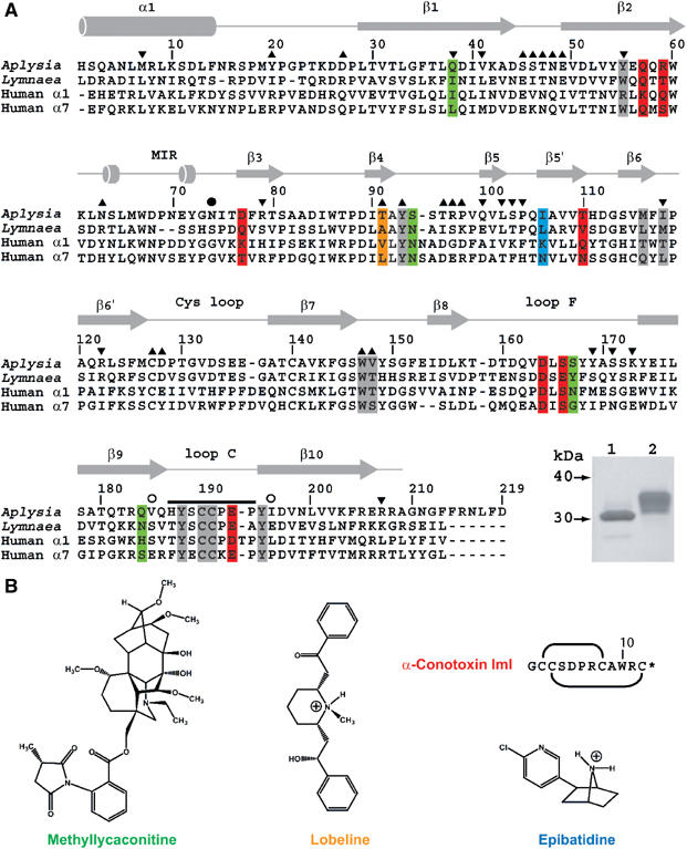
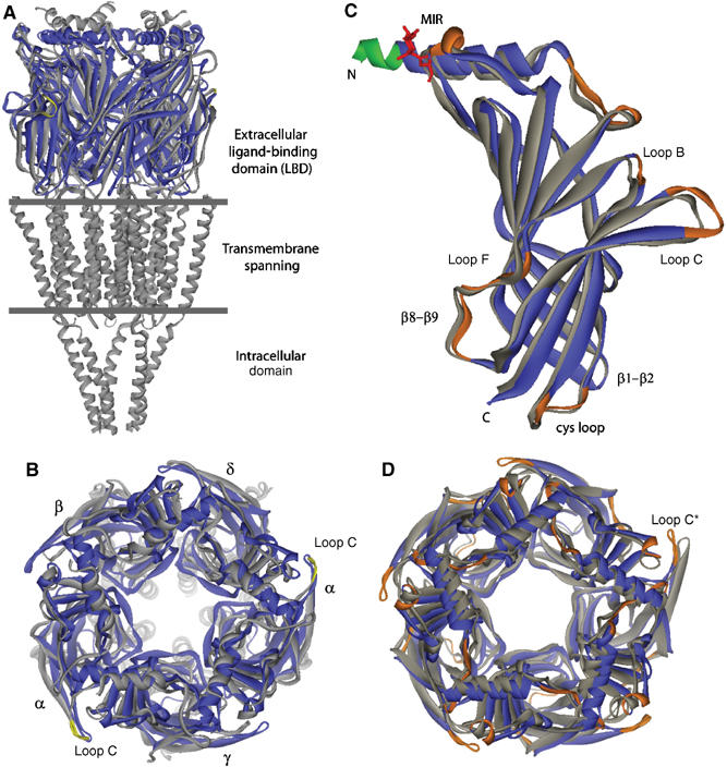
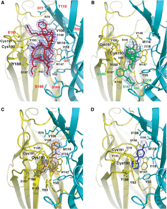
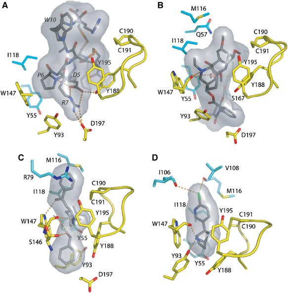
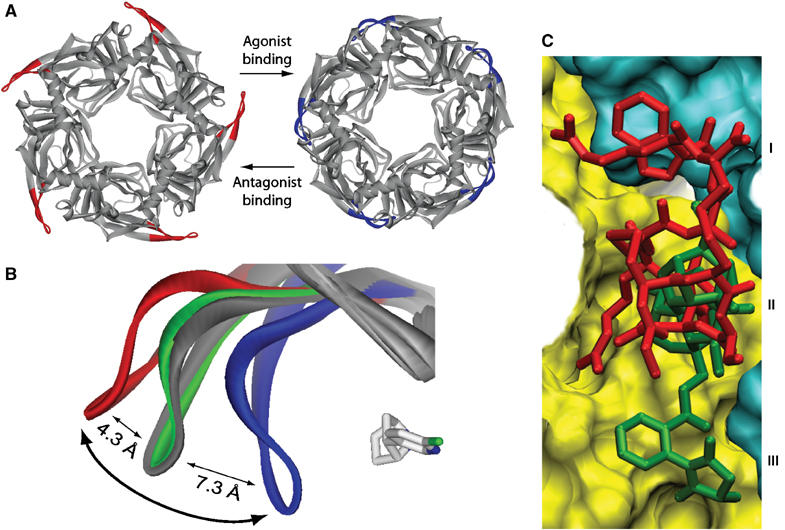
Upon ligand binding at the subunit interfaces, the extracellular domain of the nicotinic acetylcholine receptor undergoes conformational changes, and agonist binding allosterically triggers opening of the ion channel. The soluble acetylcholine-binding protein (AChBP) from snail has been shown to be a structural and functional surrogate of the ligand-binding domain (LBD) of the receptor. Yet, individual AChBP species display disparate affinities for nicotinic ligands. The crystal structure of AChBP from Aplysia californica in the apo form reveals a more open loop C and distinctive positions for other surface loops, compared with previous structures. Analysis of Aplysia AChBP complexes with nicotinic ligands shows that loop C, which does not significantly change conformation upon binding of the antagonist, methyllycaconitine, further opens to accommodate the peptidic antagonist, alpha-conotoxin ImI, but wraps around the agonists lobeline and epibatidine. The structures also reveal extended and nonoverlapping interaction surfaces for the two antagonists, outside the binding loci for agonists. This comprehensive set of structures reflects a dynamic template for delineating further conformational changes of the LBD of the nicotinic receptor.


Figures

Figure 1
Figure 1
Sequence alignment of A-AChBP. (A) Structural alignment of the subunit sequences of A-AChBP (Hansen et al, 2004) and L-AChBP (Smit et al, 2001) with those of the human α1 and α7 LBDs (LGIC database). The A-AChBP sequence reported in Celie et al (2005) differs by Val substitutions at positions 43 and 138 and a N-terminal two-residue deletion. Secondary structure elements are indicated. The bar and open circles above the A-AChBP sequence indicate the loop C tip and hinge regions, respectively. The solid circle denotes the glycosylated Asn74. Tip up and down triangles denote A-AChBP residues from the (+) and (−) faces that are within a 3.5 Å radius of interaction at the subunit interface in the apo conformation. A-AChBP residues whose side chains interact within 4.5 Å with all four ligands are on a gray background. Residues specific for the nicotinic antagonists, α-conotoxin ImI and MLA, are on a red and green background and those specific for the nicotinic agonists, LOB and EPI, on an orange and a blue background, respectively. Inset: SDS–PAGE analysis (16% gel) of A-AChBP expressed from GnTI (lane 1) and standard HEK cells (lane 2). The protein expressed in GnTI cells migrates faster and as a thinner band, indicative of a smaller size and greater homogeneity in oligosaccharide structure. (B) Schematic view of the organic ligands, MLA (the lycoctonine ring is at the top and the N-ethylpiperidine ring at the bottom), LOB and EPI. Top right: sequence and disulfide bonding of α-conotoxin ImI; the star denotes C-terminal amidation.
Figure 2
Figure 2
Overall view of the apo A-AChBP structure and structural comparisons. (A) Side view and (B) apical view of apo A-AChBP (blue) overlaid with the muscle-type nAChR (gray) (Unwin, 2005). The tip of loop C in the nAChR α subunits is colored yellow. Loop C in apo A-AChBP overlays most closely with loop C in the α subunit. (C) Side view and (D) top view of the apo A-AChBP subunit and pentamer (blue) overlaid with the Hepes-bound A-AChBP subunit and pentamer (gray) using residues 20–200 from a single and all five subunits, respectively. In apo A-AChBP, the Asn74-linked glycan is in red and the FLAG epitope that prolongs helix α1 in green. The ‘MIR' and loops where there is a large departure in position or conformation are shown in orange. In four of the five Hepes-bound A-AChBP subunits, loop C is more closed than in apo A-AChBP, whereas in the fifth subunit (star), devoid of a bound buffer, it is slightly more extended.
Figure 3
Figure 3
The A-AChBP subunit interface in the antagonist and agonist complexes. Side views of the bound antagonists (A) ImI, buried under loop C, and (B) MLA (same orientation), and of the bound agonists (C) LOB and (D) EPI in similar orientations. The 2.0–3.4 Å resolution omits 2FOFc electron density maps contoured at 1.4σ are shown in blue. The main and side chains from the (+) and (−) faces of the subunit interface are in yellow and cyan, respectively. Those side chains that interact specifically with ImI, MLA, LOB, and EPI are labeled red, green, orange, and blue, respectively.
Figure 4
Figure 4
Expanded views of the bound ligands. Hydrogen bonding of key residues for the bound (A) ImI, (B) MLA, (C) LOB, and (D) EPI, viewed from inside of the ion channel vestibule looking in a radial direction. Ligands are bound between the Cys190–Cys191 disulfide on the left and Trp148 on the right. Labels for the ImI residues are italicized. The molecular surfaces of the ligands are in gray. Subunit coloring is identical to that in Figure 3.
Figure 5
Figure 5
Conformational fit mechanism and ligand selectivity. (A) Top views of the ImI-bound (red loops C) and EPI-bound A-AChBP pentamers (blue loops C) showing the distinctive conformations for the antagonist and agonist complexes. (B) Overlay of loop C in apo A-AChBP (gray) and the ImI (red), MLA (green), and EPI complexes (blue); the bound EPI molecule is shown in light gray. The curved arrow denotes opening and closure of loop C upon antagonist and agonist binding. (C) Overlay of the A-AChBP-bound antagonists ImI (red) and MLA (green). The surfaces of the (+) and (−) faces of the subunit interface are shown in yellow and cyan, respectively. The common competitive binding site is at the center (labeled II); the two peripheral nonoverlapping sites are distinguished by ImI Trp10 (I) and the MLA N-phenyl succinimide moiety (III).

References

    1. Badio B, Daly JW (1994) Epibatidine, a potent analgetic and nicotinic agonist. Mol Pharmacol 45: 563–569 - PubMed
    1. Barlow RB, Johnson O (1989) Relations between structure and nicotine-like activity: X-ray crystal structure analysis of (−)-cytisine and (−)-lobeline hydrochloride and a comparison with (−)-nicotine and other nicotine-like compounds. Br J Pharmacol 98: 799–808 - PMC - PubMed
    1. Bergmeier SC, Ismail KA, Arason KM, McKay S, Bryant DL, McKay DB (2004) Structure activity studies of ring E analogues of methyllycaconitine. Part 2: Synthesis of antagonists to the α3β4 nicotinic acetylcholine receptors through modifications to the ester. Bioorg Med Chem Lett 14: 3739–3742 - PubMed
    1. Bourne Y, Talley TT, Hansen S B, Taylor P, Marchot P (2005) Crystal structure of a Cbtx–AChBP complex reveals essential interactions between snake α-neurotoxins and nicotinic receptors. EMBO J 24: 1512–1522 - PMC - PubMed
    1. Bouzat C, Gumilar F, Spitzmaul G, Wang HL, Rayes D, Hansen SB, Taylor P, Sine SM (2004) Coupling of agonist binding to channel gating in an ACh-binding protein linked to an ion channel. Nature 430: 896–900 - PubMed

Publication types

MeSH terms