. 2005 Oct 19;24(20):3624-34.
doi: 10.1038/sj.emboj.7600824. Epub 2005 Sep 29.

Homo- and heterodimerization of APP family members promotes intercellular adhesion

Affiliations

Homo- and heterodimerization of APP family members promotes intercellular adhesion

Peter Soba et al. EMBO J. .

Erratum in

  • EMBO J. 2006 Feb 8;25(3):653

Abstract

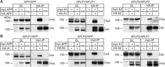
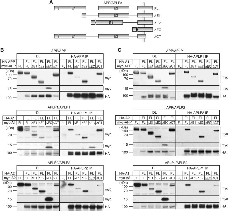
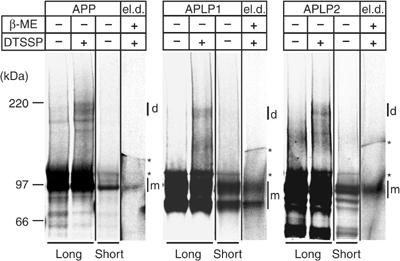
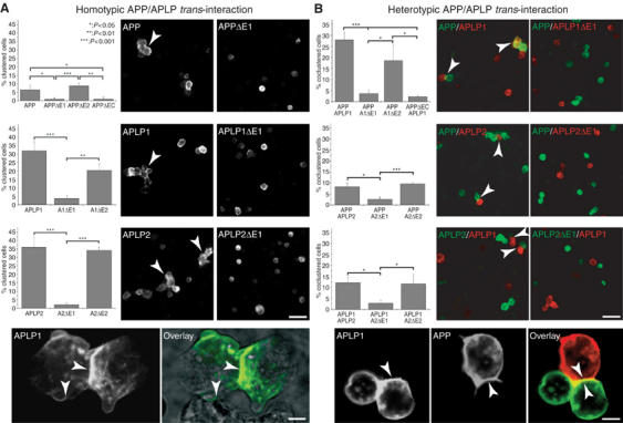
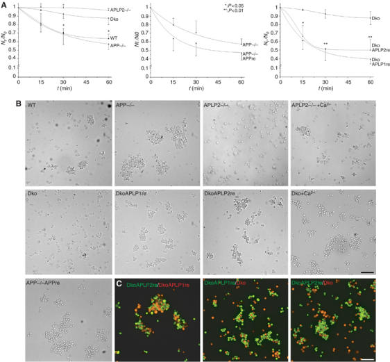
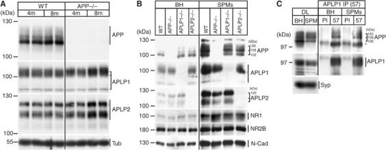
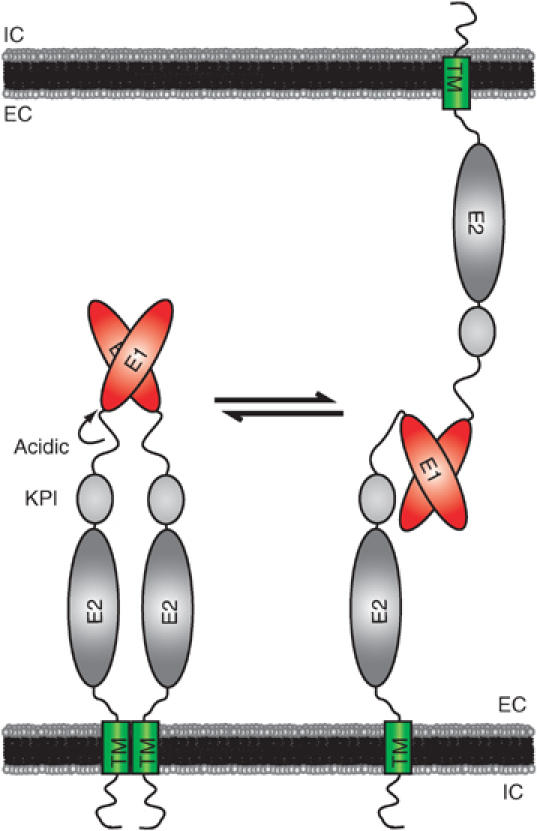
The amyloid precursor protein (APP) plays a central role in Alzheimer's disease, but its physiological function and that of its mammalian paralogs, the amyloid precursor-like proteins 1 and 2 (APLPs), is still poorly understood. APP has been proposed to form dimers, a process that could promote cell adhesion via trans-dimerization. We investigated the dimerization and cell adhesion properties of APP/APLPs and provide evidence that all three paralogs are capable of forming homo- and heterocomplexes. Moreover, we show that trans-interaction of APP family proteins promotes cell-cell adhesion in a homo- and heterotypic fashion and that endogenous APLP2 is required for cell-cell adhesion in mouse embryonic fibroblasts. We further demonstrate interaction of all the three APP family members in mouse brain, genetic interdependence, and molecular interaction of APP and APLPs in synaptically enriched membrane compartments. Together, our results provide evidence that homo- and heterocomplexes of APP/APLPs promote trans-cellular adhesion in vivo.


Figures

Figure 1
Figure 1
Homo- and heterointeraction of APP family proteins. (A) Homointeraction of myc- and HA-tagged APP, APLP1, and APLP2. Pairs of myc- and HA-tagged APP (myc-APP and HA-APP), APLP1 (myc-A1 and HA-A1), or APLP2 (myc-A2 and HA-A2) were expressed in COS7 cells. In all, 1/25 of each lysate was used as an input control (DL). HA-tagged APP/APLPs were immunoprecipitated and immunoblotted for myc- and HA-tagged constructs. As controls, cells transfected with the corresponding myc-tagged APP family member or postlysis mixtures (lanes ‘mix') of separately transfected myc- and HA-tagged APP/APLPs were immunoprecipitated. (B) Heterointeraction of myc- and HA-tagged APP, APLP1, and APLP2. Heterotypic pairs of myc- and HA-tagged APP, APLP1, or APLP2 were transfected into COS7 cells as indicated. In all, 1/25 of the each lysate was loaded as input control (DL). Myc-tagged APP/APLPs were immunoprecipitated and immunoblotted for myc- and HA-tagged APP/APLPs. Controls were performed as above.
Figure 2
Figure 2
Mapping of APP/APLP homo- and heterointeraction. (A) Schematic drawing of APP/APLP constructs used. HA- and myc-tagged APP/APLP constructs lacking either the E1 domain (ΔE1), the E2 domain (ΔE2), the entire ectodomain (ΔEC), or the cytoplasmic domain (ΔCT) were analyzed. (B) Homointeraction of full-length HA-tagged APP, APLP1, and APLP2 with different deletion constructs in COS7 cells. HA-tagged APP (HA-APP FL) was coexpressed with vector only, myc-tagged APPΔE1 (ΔE1), APPΔE2 (ΔE2), APPΔEC (ΔEC), or APPΔCT (ΔCT). In all, 1/25 of each lysate was used for the DL. HA-tagged APP was immunoprecipitated from cell extracts and immunoblotted for myc- and HA-tagged constructs. The identical setup was used for APLP1 (HA-A1) and APLP2 (HA-A2) homointeraction with the corresponding deletion constructs. Lower levels of APLP2ΔCT interaction are due to lower expression levels of this construct. (C) Heterointeraction of APP, APLP1, and APLP2 with different deletion constructs in COS7 cells. As for the mapping of homointeractions, a HA-tagged APP family member was coexpressed with vector only or different APP/APLP deletion constructs and immunoprecipitated (as indicated). In all, 1/25 of each lysate was used as an input control (DL).
Figure 3
Figure 3
Dimerization of APP family proteins at the cell surface. [35S-Met]-labeled COS7 cells expressing APP, APLP1, or APLP2 were incubated either with or without the membrane-impermeable crosslinker DTSSP as indicated. APP/APLPs were immunoprecipitated with anti-APP (22734), anti-APLP1 (57), or anti-APLP2 (D2-II) antibodies, respectively. The samples were denatured without β-mercaptoethanol (β-ME) and analyzed on 3–8% Tris-acetate gels by autoradiography. Long and short exposures are shown as indicated to visualize crosslinked dimeric (d) or the monomeric (m) forms of APP/APLPs, respectively. Longer exposures show the crosslinked dimers compared to control cells without crosslinker. The crosslinking products (d) were subsequently extracted from the gel, denatured under reducing conditions (el.d.: eluted dimers), and analyzed on 3–8% Tris-acetate gels. The asterisks indicate unspecific signals present in all samples and controls.
Figure 4
Figure 4
Homo- and heterotypic intercellular interactions of APP, APLP1, and APLP2. (A) Quantification and immunostainings of homotypic S2 cell clusters (indicated by arrows) expressing APP/APLPs or the corresponding deletion constructs. The percentage of clustered transfected cells from at least three independent experiments is shown for the different constructs as indicated (n⩾3, ±s.d., t-test, scale bar=20 μm). Lower panel: confocal analysis of APLP1 expressing S2 cells stained with an anti-myc antibody (APLP1) and overlaid with the transmission image (scale bar=3 μm). (B) Quantification and immunostainings of heterotypic cell contacts (indicated by arrows) of mixed pools of S2 cells expressing APP/APLPs or the corresponding extracellular deletion constructs as indicated. Direct heterotypic cell contacts were quantified from at least three independent experiments, and is given as the percentage of total transfected cells (n⩾3, ±s.d., t-test, scale bar=20 μm). Lower panel: confocal analysis of immunostained mixed pools of S2 cells expressing APLP1 (anti-myc) and APP (40090, scale bar=3 μm).
Figure 5
Figure 5
APP family proteins are required for cell adhesion. (A) Quantitative cellular aggregation of WT, APP knockout (APP−/−), APLP2 knockout (APLP2−/−), or APP/APLP2 double-knockout MEF cells (Dko). MEF cells were aggregated in suspension under calcium- and magnesium-free conditions for 1 h at 80 r.p.m. The number of particles (single cells and cell clusters) was counted at the indicated time points (Nt). The relative decrease in particle counts compared to t=0 (Nt/N0) is shown over time as a measure of aggregation of the different MEF cells (n⩾3, ±s.d., t-test). APP−/− MEFs were compared with APP retransfected cells (APP−/−APPre), and APLP1 (DkoAPLP1re) or APLP2 (DkoAPLP2re) rescued Dko cells were compared with parental Dko cells. (B) Typical micrographs of the different MEF lines after 60 min of aggregation. APLP2−/− and Dko cells were additionally aggregated in the presence of 1 mM Ca2+ to induce cadherin-mediated adhesion (scale bar=100 μm). (C) Equal amounts of differentially labeled Dko, DkoAPLP1re, and DkoAPLP12re cells (as indicated) were coaggregated for 60 min and analyzed by fluorescence microscopy (scale bar=100 μm).
Figure 6
Figure 6
APP/APLPs form heterocomplexes in vivo. Brain extracts from WT, APP−/−, APLP1−/−, and APLP2−/− mice were subjected to immunoprecipitation and SDS–PAGE. In all, 20 μg of total extracts (DL) was loaded for comparison. (A) APP/APLP1 interaction was probed by immunoprecipitating with the anti-APLP1 antibody 57 and Western blot detection of APP (22C11), APLP1 (CT-11), and β-tubulin III (Tub). Note that only mature higher-molecular-weight forms of APP were coimmunoprecipitated with APLP1. The apparent size of the different APP bands is indicated. (B) APP/APLP2 interaction was probed by immunoprecipitating with the anti-APP antibody 22734 and Western blot detection of APLP2 (D2-13), APP (22C11), and β-tubulin III (Tub). The apparent size of the different APLP2 bands is indicated. (C) APLP1/APLP2 interaction was probed by immunoprecipitating with the anti-APLP1 antibody 57 and Western blot detection of APLP2 (D2-13), APLP1 (CT-11), and β-tubulin III (Tub).
Figure 7
Figure 7
Accumulation of APLPs in APP−/− mouse brain and synaptic enrichment and interaction. (A) In all, 20 μg of total mouse brain lysates from 4- (4 m) or 8-months- (8 m) old WT and APP−/− mice were subjected to SDS–PAGE and immunoblotted for APP, APLP1, and APLP2, and β-tubulin III (Tub). The size of the different APP/APLP bands is indicated by brackets. Note that that the highest APLP1 band is specifically accumulating in APP−/− mice compared to WT (size is indicated by brackets and white lines). (B) Synaptic plasma membranes (SPMs) were prepared from BH of WT, APP−/−, APLP1−/−, and APLP2−/− mice. From each fraction, 20 μg of total protein was subjected to SDS–PAGE and immunoblotted with antibodies against APP (22C11), APLP1 (CT-11), and APLP2 (D2-II). Enrichment of synaptic compartments was confirmed with NMDA receptor 1 (NR1) and 2B (NR2B), and N-cadherin antibodies (N-Cad). (C) Total extracts (BH) and SPM preparations from WT mouse brains were directly loaded to the gel (DL) or immunoprecipitated either with PI or an anti-APLP1 antibody (57), followed by Western blot detection of APP (22C11), APLP1 (CT-11), and synaptophysin (Syp). DLs of BH and SPMs are shown in comparison. The asterisk indicates a signal of the antibody heavy chain.
Figure 8
Figure 8
A model for cis- and trans-interaction of APP family proteins. The schematic model of APP/APLP domain organization and interaction is shown. The N-terminal E1 domain is linked to a highly flexible acidic region, followed by the alternatively spliced Kunitz-type protease inhibitor (KPI) domain (for APP and APLP2), the E2 domain, the juxtamembrane/TM region, and the cytosolic domain. Based on our results, we suggest that APP family proteins are capable of forming lateral and adhesive dimers in homo- and heterotypic fashions. The E1 domain is crucial for both cis- and trans-interactions, while the TM region could additionally contribute to lateral dimerization.

Similar articles

Cited by

References

    1. Aguzzi A, Haass C (2003) Games played by rogue proteins in prion disorders and Alzheimer's disease. Science 302: 814–818 - PubMed
    1. Annaert W, De Strooper B (2002) A cell biological perspective on Alzheimer's disease. Annu Rev Cell Dev Biol 18: 25–51 - PubMed
    1. Beher D, Elle C, Underwood J, Davis JB, Ward R, Karran E, Masters CL, Beyreuther K, Multhaup G (1999) Proteolytic fragments of Alzheimer's disease-associated presenilin 1 are present in synaptic organelles and growth cone membranes of rat brain. J Neurochem 72: 1564–1573 - PubMed
    1. Beher D, Hesse L, Masters CL, Multhaup G (1996) Regulation of amyloid protein precursor (APP) binding to collagen and mapping of the binding sites on APP and collagen type I. J Biol Chem 271: 1613–1620 - PubMed
    1. Coulson EJ, Paliga K, Beyreuther K, Masters CL (2000) What the evolution of the amyloid protein precursor supergene family tells us about its function. Neurochem Int 36: 175–184 - PubMed

Publication types

MeSH terms