Comparative Study
. 2005 Dec;234(4):878-91.
doi: 10.1002/dvdy.20565.

SOX7 and SOX18 are essential for cardiogenesis in Xenopus

Affiliations
Comparative Study

SOX7 and SOX18 are essential for cardiogenesis in Xenopus

Chi Zhang et al. Dev Dyn. 2005 Dec.

Abstract

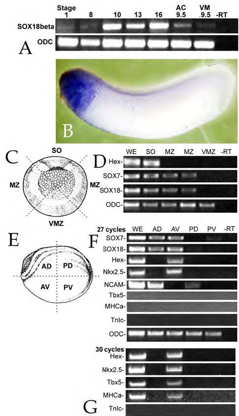
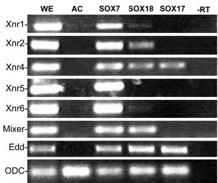
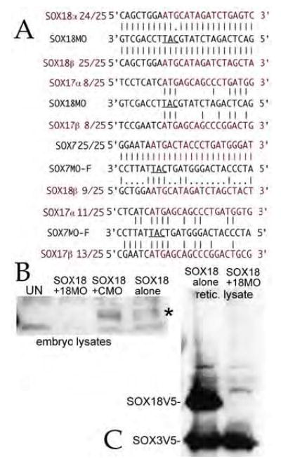
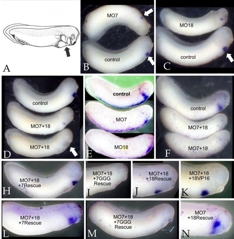
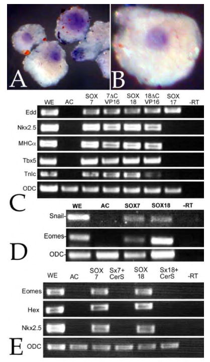
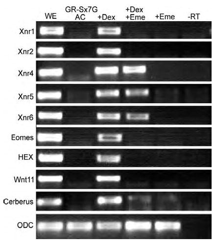
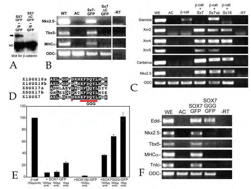
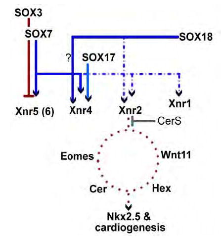
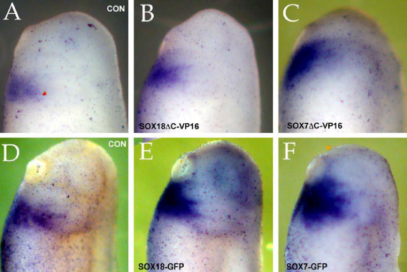
Early in vertebrate development, endodermal signals act on mesoderm to induce cardiogenesis. The F-type SOXs SOX7 and SOX18beta are expressed in the cardiogenic region of the early Xenopus embryo. Injection of RNAs encoding SOX7 or SOX18beta, but not the related F-type SOX, SOX17, leads to the nodal-dependent expression of markers of cardiogenesis in animal cap explants. Injection of morpholinos directed against either SOX7 or SOX18mRNAs lead to a partial inhibition of cardiogenesis in vivo, while co-injection of SOX7 and SOX18 morpholinos strongly inhibited cardiogenesis. SOX7 RNA rescued the effects of the SOX18 morpholino and visa versa, indicating that the proteins have redundant functions. In animal cap explants, it appears that SOX7 and SOX18 act indirectly through Xnr2 to induce mesodermal (Eomesodermin, Snail, Wnt11), organizer (Cerberus) and endodermal (endodermin, Hex) tissues, which then interact to initiate cardiogenesis. Versions of SOX7 and SOX18 with their C-terminal, beta-catenin interaction domains replaced by a transcriptional activator domain failed to antagonize beta-catenin activation of Siamois, but still induced cardiogenesis. These observations identify SOX7 and SOX18 as important, and previously unsuspected, regulators of cardiogenesis in Xenopus.


Figures

Figure 1
Figure 1. Expression and activity of SOX18β
A: Fertilized eggs were analyzed by RT-PCR for SOX18 RNA at various stages. A weak signal was observed in pre-MBT embryos, but strong expression was detected by stage 10. When animal caps and vegetal masses were analyzed at stage 9.5, SOX18β mRNA appeared to be present at similar levels in both regions. Ornithine decarboxylase (ODC) was used as a control for RNA quality throughout. B: In situ hybridization staining with an anti-sense SOX18β probe revealed expression primarily in the anterior region of stage 30 embryos – at earlier stages no consistent, regionspecific staining was observed; no staining was observed using a sense probe (data not shown). C: To define the regional distribution of SOX7 and SOX18 in the gastrula (stage 10/11) embryos, such embryos were dissected into four regions: Spemann Organizer/Dorsal marginal zone (“SO”), lateral marginal zones (“MZ”), and ventral marginal zone (“VZ”). The schematic shows these regions. D: RNA was isolated from the various regions and used for RT-PCR analysis (30 cycles); SOX7 and SOX18 RNAs were found in the SO and MZ, but not in the VZ region; Hex RNA was restricted to the SO region. ODC was found to be uniformly distributed in all four regions. E: A similar analysis was carried out with neurula stage embryos (stage 16/17); these embryos were dissected into anterior dorsal (“AD”), anterior ventral (“AV”), posterior dorsal (“PD”) and posterior ventral (“PV”) regions and RT-PCR analysis was performed. F: Using 27 cycles of amplification, we found SOX7 and SOX18 RNA in both anterior regions, but little if any in the posterior regions. Hex and Nkx2.5 RNAs were found in the AV region only, while NCAM RNA (a marker of neural specification) was found in the two dorsal regions (AD and PD) but not in ventral regions. G: When 30 cycles of PCR were used, Tbx5 and MHCa, but not TnIc RNAs could be detected in the anterior ventral region of the embryo. Images in parts C and E are modified from Nieuwkoop & Faber (1976).
Figure 2
Figure 2. SOX18 induction of Nodal-related genes in animal caps
To compare the activity of SOX18 to SOX7 and SOX17 in the animal cap system, fertilized eggs were injected with RNAs encoding SOX7-GFP, mtSOX17β-GFP or SOX18β- GFP (0.5 ng/embryo). At stage 8, animal caps were prepared, cultured for approximately 4 hours (when control embryos had reached stage 10/11) and then analyzed by RT-PCR (Unless otherwise noted, this is the “standard” animal cap assay used throughout this work). As reported previously, SOX7-GFP induced the expression of the five mesoderminducing nodal-related genes Xnr1, Xnr2, Xnr4, Xnr5 and Xnr6, as well as Mixer and Endodermin (Edd), while SOX17β-GFP induce the expression of Xnr4 and Edd, but not Mixer. SOX18β-GFP induced weak expression of Xnr1, and stronger expression of Xnr2, Xnr4, Mixer and Edd.
Figure 3
Figure 3. SOX7 and SOX18 morpholinos
A: The sequence of the morpholinos against SOX7 and SOX18, their alignment with their target sequences, and the analogous regions of the other F-type SOX mRNAs are shown. The ability of the SOX7 morpholino to inhibit SOX7 RNA translation has been established previously (Zhang et al., 2005). B: To test the specificity of the SOX18 morpholino, fertilized eggs where injected with 0.5 ng/embryo utr-SOX18β-V5 RNA either alone or in the presence of control (CMO) or SOX18 (18MO) morpholinos (15 ng/embryo). At stage 8/9 embryonic lysates were generated and analyzed by SDS-PAGE/immunoblot using a monoclonal antiV5 antibody. The accumulation of SOX18β-V5 polypeptides (marked “*”) was blocked by the SOX18 morpholino, but unaffected by the control morpholino. C: Rabbit reticulocyte extracts (25 μL final volume) were programmed with 2 μg each of utr-SOX18β-V5 and SOX3-V5 RNAs either alone or together with 80ng SOX18 morpholino. The reactions were then analyzed by SDS-PAGE and immunoblot using the monoclonal antiV5 antibody. The SOX18 morpholino completely inhibited the accumulation of SOX18-V5 polypeptide, but had no effect on SOX3V5 polypeptide accumulation.
Figure 4
Figure 4. Inhibition of cardiogenesis by SOX7 and SOX18 morpholinos
Fertilized eggs were injected at animal and vegetal sites with a total of 30 ngs of morpholino and examined at stage ~30. A: A schematic of a stage 30 embryo showing the location of the cardiogenic region (“arrow”)(Image from Nieuwkoop & Faber, 1976). In situ hybridization using antisense RNAs directed against either MHCα (B-D) or Nkx2.5 (E,F). Both the SOX7 morpholino (MO7)(B,E) and the SOX18 morpholino (MO18) (C,E) reduced the size and intensity of the MHCα and Nkx2.5 staining domains. A combination of both morpholinos (15+15 ng/embryo)(D,F) produced a more complete suppression of MHCα and Nkx2.5 staining (see also Table 2). Rescue studies: To confirm the specificity of the morpholino effects, MO7+MO18-injected embryos were injected with RNAs (0.5 ng/embryo) encoding either altSOX7-GFP (H,L), SOX7GGG-GFP (I,M), SOX18β-GFP (J,N), or mtSOX18βΔC-VP16 (18VP16)(K); at stage 30 the embryos were stained in situ for MHCα (H-K) or Nkx2.5 (L-N). SOX7, SOX18, and mtSOX18βΔC-VP16 rescued the effects of the combined morpholinos, whereas SOX7GGG-GFP did not.
Figure 5
Figure 5. Induction of cardiogenesis in animal caps
To determine whether SOX7 induced markers of cardiogenesis, fertilized eggs were injected with RNA encoding SOX7-GFP (0.5ng/embryo); animal caps were prepared at stage 8 and analyzed by in situ hybridization when control embryo had reached stage 30. Both Nkx2.5 (A) or MHCα (B) were expressed in discrete domains, one per animal cap; no expression of either gene was observed in cap derived from uninjected embryos (data not shown). C: A similar analysis was carried using RT-PCR. Fertilized eggs were injected with RNA (0.5 ng/embryo) encoding either SOX7-GFP (SOX7), mtSOX7ΔC-VP16 (7ΔCVP16), SOX18β-GFP (SOX18), mtSOX18ΔC-VP16 (18ΔCVP16), or mtSOX17β-GFP (SOX17). Animal caps were prepared and analyzed when controls reached stage 25. Each of the injected RNAs induced the expression of the endodermal marker Edd, but only SOX7 or SOX18 constructs induced markers of cardiogenesis, Nkx2.5, MHCα Tbx5 and TnIc. D: In a similar study, SOX7-GFP and SOX18β-GFP induced expression of the mesodermal markers Snail and Eomesodermin (Eomes). E: The SOX7-GFP and SOX18β-GFP induction of Eomes, Hex and Nkx2.5 expression was blocked by the coinjection of RNA encoding the nodal inhibitor CerS (0.2 ng/embryo).
Figure 6
Figure 6. Direct versus indirect targets of SOX7 regulation
Fertilized eggs were injected with RNA encoding GR-SOX7-GFP (0.5 ng/embryo). Animal caps were treated in four different ways: either left in standard media (AC) with 1% ethanol (the carrier for the dexamethasone), incubated with 20 μM dexamethasone (+DEX), pretreated with 100 μg/ml emetine for 30 minutes and then incubated with 20mM dexamethasone and 100 μg/ml emetine (+DEX +Eme), or incubated with 100 μg/ml emetine alone (+Eme). In contrast to cycloheximide (see Sinner et al 2004), emetine treatment had no apparent effect on the expression of any of the genes examined in the absence of dexamethasone. In the presence of dexamethasone, the Xnrs were induced, along with Eomes, Hex, Wnt11 and Cerberus; of these genes, Xnr4, Xnr5 and Xnr6 were expressed in the presence of both dexamethasone and emetine, indicating that they are direct targets of SOX7 regulation.
Figure 7
Figure 7. SOX7 antagonism of b-catenin signaling and cardiogenic ability
A: Fertilized eggs were injected with RNA encoding either SOX7-GFP (Sx7GFP) or SOX7ΔC-GFP (Sx7ΔC-GFP)(0.5 ng/embryo); at stage 9/10 embryo lysates were prepared and immunoprecipitated with a rabbit antiGFP antibody. Immunoprecipitates where analyzed by SDS-PAGE and immunoblot using a rabbit anti-β-catenin antibody. β-catenin was coprecipitated with SOX7-GFP (arrow), but little or no β-catenin co-precipitated with SOX7ΔC-GFP (immunoglobulin heavy chain marked “HC”). Immunoblot with antiGFP antibody of embryonic lysates indicated that both SOX7-GFP and SOX7ΔC-GFP polypeptides accumulated to similar extents (data not shown). B: In an animal cap assay, injection of RNA (0.5 ng/embryo) encoding SOX7ΔC-GFP failed to induce Nkx2.5, Tbx5 or MHCα expression (animal caps analyzed when control embryos reached stage 25). C: Injection of RNA encoding a mutationally stabilized form of β-catenin (mt-ΔG-β-catenin) induces the expression of Siamois in animal caps. Fertilized eggs were injected with RNA encoding mt-ΔG-β-catenin (50 pg/embryo) alone or together with RNAs encoding SOX7-GFP, mt-SOX7ΔC-VP16 or SOX18β-GFP (500 pg/embryo); animal caps were prepared at stage 8 and analyzed when control embryos reached stage 10/11. β-catenin induced expression of Siamois; both SOX7-GFP and SOX18β-GFP suppressed β-catenin-induced Siamois expression, and induced expression of Xnr2, Xnr4, Cerberus and Nkx2.5; SOX7-GFP also induced Xnr5 expression. SOX7ΔC-VP16 failed to suppress β-catenin-induced Siamois expression, but induced Xnr2, Xnr4, Xnr5, Cerberus and Nkx2.5 expression. D: The SOX7GGG-GFP construct is analogous to the SOX17G3 construct generated and characterized by Sinner et al (2004). In animal caps assayed at stage 25, SOX7GGG-GFP (0.5 ng/embryo) induced the expression of Edd, but not Nkx2.5, Tbx5, MHCα or TnIc. E: To examine the ability of SOX7-GFP, SOX7GGG-GFP and SOX18β-GFP to inhibit β-catenin activation of the OT reporter, fertilized eggs were injected with OT and pTK-Renilla DNAs (20 pgs each/embryo) together with RNAs encoding mt-ΔG-β-catenin (50 pg/embryo) and varying amounts of SOX RNAs; animal caps were analyzed when control embryos reached stage 10/11. SOX7-GFP and SOX18β-GFP RNAs inhibited of β-catenin activation of OT; SOX7GGG-GFP was a much less active antagonist of β-catenin in this assay.
Figure 8
Figure 8. SOX7, SOX18 and the cardiogenic pathway
In this diagram, direct interactions are indicated by solid lines, indirect interactions by dotted lines. SOX17 induces Xnr4, but fails to induce cardiogenesis in animal caps, whereas SOX7 and SOX18 induce both Xnr2 and Xnr4 and induce cardiogenesis. The presence of the nodal inhibitor CerS inhibits SOX7 and SOX18-induced cardiogenesis. How other direct and indirect targets of SOX7 and SOX18 interact with “downstream” targets of Xnr-regulation (e.g. Eomes, Wnt11, Cerberus and Hex) remains unclear - nevertheless, the end result in the activation of Nkx2.5 and other cardiac markers by SOX7 and SOX18.
Supplemental figure
Supplemental figure
Fertilized eggs were injected with either SOX18βΔC-VP16myc (B), SOX7ΔC-VP16myc (C), SOX18β-GFP (E) or SOX7-GFP (F) RNAs (0.5 ng/embryo). At stages between 25 (A-C) and 30 (D-F) the embryos were stained in situ for Nkx2.5. Compared to uninjected controls (A,D), both GFP and ΔCVP16myc forms of SOX7 and SOX18 induced an apparent increase in the extent and intensity of the Nkx2.5 expression domain.

Similar articles

Cited by

References

    1. Agius E, Oelgeschlager M, Wessely O, Kemp C, De Robertis EM. Endodermal Nodal-related signals and mesoderm induction in Xenopus. Development. 2000;127:1173–83. - PMC - PubMed
    1. Ariizumi T, Kinoshita M, Yokota C, Takano K, Fukuda K, Moriyama N, Malacinski GM, Asashima M. Amphibian in vitro heart induction: a simple and reliable model for the study of vertebrate cardiac development. Int J Dev Biol. 2003;47:405–10. - PubMed
    1. Aybar MJ, Nieto MA, Mayor R. Snail precedes slug in the genetic cascade required for the specification and migration of the Xenopus neural crest. Development. 2003;130:483–94. - PubMed
    1. Bouwmeester T, Kim S, Sasai Y, Lu B, De Robertis EM. Cerberus is a head-inducing secreted factor expressed in the anterior endoderm of Spemann’s organizer. Nature. 1996;382:595–601. - PubMed
    1. Brannon M, Kimelman D. Activation of Siamois by the Wnt pathway. Dev Biol. 1996;180:344–7. - PubMed

Publication types

MeSH terms

LinkOut - more resources