. 2005 Nov 25;280(47):39474-84.
doi: 10.1074/jbc.M504672200. Epub 2005 Sep 29.

Rac1 is essential for platelet lamellipodia formation and aggregate stability under flow

Affiliations

Rac1 is essential for platelet lamellipodia formation and aggregate stability under flow

Owen J T McCarty et al. J Biol Chem. .

Abstract

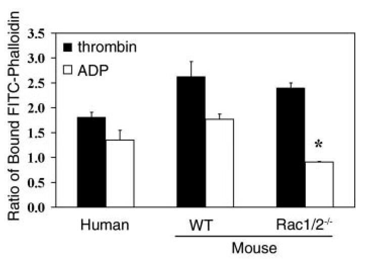
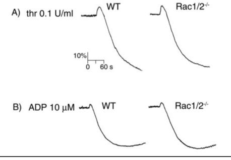
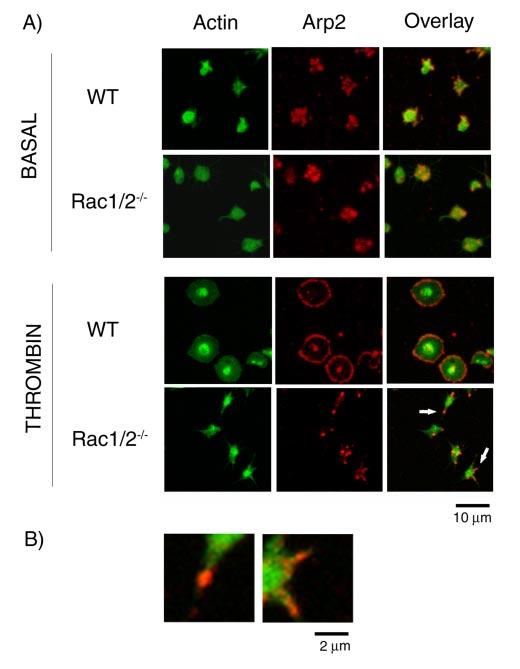
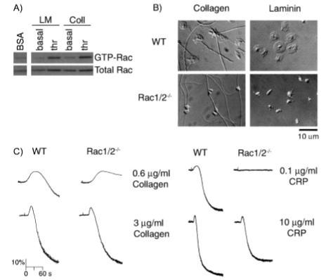
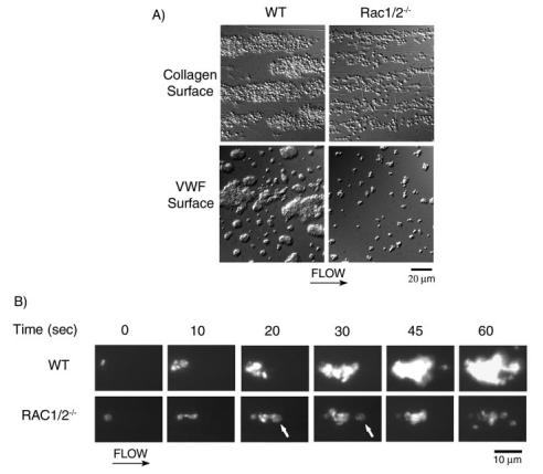
The role of Rac family proteins in platelet spreading on matrix proteins under static and flow conditions has been investigated by using Rac-deficient platelets. Murine platelets form filopodia and undergo limited spreading on fibrinogen independent of Rac1 and Rac2. In the presence of thrombin, marked lamellipodia formation is observed on fibrinogen, which is abrogated in the absence of Rac1. However, Rac1 is not required for thrombin-induced aggregation or elevation of F-actin levels. Formation of lamellipodia on collagen and laminin is also Rac1-dependent. Analysis of platelet adhesion dynamics on collagen under flow conditions in vitro revealed that Rac1 is required for platelet aggregate stability at arterial rates of shear, as evidenced by a dramatic increase in platelet embolization. Furthermore, studies employing intravital microscopy demonstrated that Rac1 plays a critical role in the development of stable thrombi at sites of vascular injury in vivo. Thus, our data demonstrated that Rac1 is essential for lamellipodia formation in platelets and indicated that Rac1 is required for aggregate integrity leading to thrombus formation under physiologically relevant levels of shear both in vitro and in vivo.


Figures

FIGURE 1.
FIGURE 1.
Rac isoforms present in murine and human platelets (plt). Equal amounts of human and murine platelet lysates were analyzed for Rac expression using noncross-reactive Rac1 (A) or Rac2 (B) antibodies. Lysates from wild-type (WT) and Rac2-/-thymus were included as a positive control.
FIGURE 2.
FIGURE 2.
Effects of agonists on Rac1 activation in platelets both in suspension and on a fibrinogen surface. Equal numbers of washed human (A) or murine (B) platelets (5 × 10⁸/ml) were stimulated with thrombin (thr; 1 unit/ml) or ADP (10μM) in the absence or presence of ADP scavenger apyrase (2 units/ml) for 60 s before lysis. Subsequently platelet lysates were incubated with agarose beads coupled with GST-PBD fusion proteins to bind GTP-bound forms of Rac1. Total lysates or material binding to the GST-PBD beads were solubilized, separated by SDS-PAGE, and detected by immunoblotting with an anti-Rac1 monoclonal antibody. In separate experiments, washed human (C) and murine (D) platelets (5 × 10⁸/ml) were added to BSA- or fibrinogen (FG)-coated dishes in the absence or presence of thrombin (1 unit/ml) and incubated for 10 min. Dishes coated with fibrinogen were washed twice to remove nonadherent cells. Platelets adherent to fibrinogen or in suspension over BSA were lysed, and Rac activity was determined as described above. Each panel depicts one experiment representative of three independent experiments.
FIGURE 3.
FIGURE 3.
Quantitation of F-actin in human and murine platelets responding to various stimuli. Purified human and wild-type (WT) or Rac1-/-Rac2-/-murine platelets were activated with 1 unit/ml thrombin or 10 μM ADP for 60 s. Samples were then fixed with 3.7% formaldehyde, permeabilized with 0.1% Triton X-100 containing 2 μM FITC-phalloidin, and analyzed in a fluorometer as described. Results are expressed as the ratio between the fluorescence of activated cells versus resting cells. Values are mean ± S.D. (n = 2 performed in triplicate). *,p < 0.01 with respect to wild-type platelets.
FIGURE 4.
FIGURE 4.
Platelets from Rac1/Rac2-deficient mice have normal aggregation responses. A, washed platelets (2 × 10⁸/ml) from wild-type (WT) and Rac1-/-Rac2-/-mice were stimulated with 0.1 unit/ml thrombin and the change in optical density indicative of aggregation recorded. B, heparinized PRP (2 × 10⁸/ml) from wild-type and Rac1/Rac2-deficient mice was stimulated with 10 μM ADP, and the aggregation was recorded as described above. One representative experiment of three separate trials is depicted.
FIGURE 5.
FIGURE 5.
Spreading of human and murine platelets on fibrinogen. Purified human and wild-type (WT) or Rac1-/-Rac2-/-murine platelets (2 × 10⁷/ml) were placed on coverslips coated with fibrinogen for 45 min and imaged using DIC microscopy. Platelets were treated with apyrase (2 units/ml) and indomethacin (10 μM) in the absence or presence of 1 unit/ml thrombin. In separate experiments, platelets were treated with 10 μM ADP in the absence of inhibitors of secondary mediators. Results are representative of at least three experiments.
FIGURE 6.
FIGURE 6.
Real time imaging of platelet spreading on fibrinogen. Purified murine platelets (2 × 10⁷/ml) were exposed to a fibrinogen-coated surface in the presence of apyrase (2 units/ml) and indomethacin (10 μM) and observed in real time using DIC microscopy. Representative morphology of a single wild-type (WT)(A) and (B) Rac1-/-Rac2-/- murine platelet spreading on fibrinogen in the absence or presence of thrombin (1 units/ ml). Corresponding videos 1 and 2 can be found in the supplemental material. C, the mean surface area of wild-type and Rac1-/-Rac2-/- murine platelets in the absence (filled boxes) or presence of thrombin (filled circles) or ADP (open triangles) was quantitated at the indicated time points using ImageJ as described under “Experimental Procedures.” ADP stimulation was performed in the absence of inhibitors of secondary mediators. One experiment representative of 3-5 is depicted, and values are mean ± S.E. of at least 15 cells.
FIGURE 7.
FIGURE 7.
Location of Arp2/3 in spread platelets. A, purified wild-type (WT) and Rac1/ 2-/-murine platelets (2 × 10⁷/ml) were exposed to a fibrinogen-coated surface in the presence of apyrase (2 units/ml) and indomethacin (10 μM) with or without 1 unit/ml thrombin for 45 min. Cells were fixed with 3.7% paraformaldehyde containing 0.2% Triton and stained for actin filaments (FITC-phalloidin; column 1) and Arp2/3 (rhodaminp34; column 2) before examination by confocal microscopy. B, higher magnification of the two filopodia indicated by an arrow in A. Images are representative pictures from three experiments.
FIGURE 8.
FIGURE 8.
Spreading of murine platelets on collagen and laminin. A, effects of platelet spreading on collagen and laminin on Rac activation. Washed murine platelets (5 × 10⁸/ml) were added to BSA-, collagen (Coll)-, or laminin (LM)-coated dishes in the presence of apyrase (2 units/ml) and indomethacin (10 μM) and incubated for 10 min at 37 °C. Where indicated, platelets were pretreated with thrombin (thr; 1 units/ml). Dishes were washed twice to remove nonadherent cells. Platelets adherent to either collagen or laminin or in suspension over BSA were lysed, and Rac activity was determined as described in Fig. 2. B, purified wild-type (WT), and Rac1-/-Rac2-/-, murine platelets (2 × 10⁷/ml) were placed on coverslips coated with either collagen or laminin in the presence of apyrase (2 units/ml) and indomethacin (10 μM) for 45 min before being fixed and imaged using DIC microscopy. C, aggregation of platelets from wild-type (WT) and Rac1/Rac2-deficient mice was measured as described in the legend to Fig. 4. One experiment representative of three is depicted.
FIGURE 9.
FIGURE 9.
Role of Rac in platelet adhesion and aggregate stability on collagen under flow. A, mouse blood anticoagulated with D-phenyl-alanyl-1-prolyl-1 arginine chloromethyl ketone and heparin was perfused through a collagen-coated microslide at a shear rate of 1000 s-1 for 4 min followed by modified Tyrode buffer for 3 min to remove nonadherent cells. In separate experiments, mouse blood was anticoagulated with sodium citrate and perfused over VWF/thrombin-coated microslides as described above. Subsequently, slides were fixed and visualized using DIC microscopy. Representative images of platelet adhesion from wild-type (left panel) and Rac1-/-Rac2-/- (right panel) mice are shown. B, alternatively, mouse blood was fluorescently labeled with DiOC6 prior to perfusion over collagen as described above. A representative time course for both wild-type (WT) (top panel) and Rac1-/-Rac2-/- (bottom panel) platelet accumulation on collagen is shown. Note the Rac1-/-Rac2-/- platelet embolization as indicated by the arrowhead at 20 and 30 s. Direction of flow is from left to right. Images are representative of three experiments.
FIGURE 10.
FIGURE 10.
Role of Rac in thrombus formation in vivo. A, thrombus formation over time in wild-type (top) and Rac1-/- (bottom) mice. Platelets were labeled in vivo with Alexa 488-conjugated goat anti-rat antibody bound to rat anti-CD41 antibody. The Alexa 488 fluorochrome image, collected digitally and presented as a green pseudocolor, is merged with the bright field image. Blood flow is from right to left in each image, as depicted by an arrow. Lines approximating the walls of the arteriole are shown in the 1st image of the series. B, kinetics of platelet accumulation. Each curve represents the median integrated platelet fluorescence for wild-type (WT) (8 thrombi; n = 2) and Rac1-/-(16 thrombi; n = 3) mice as a function of time after injury. C, corresponding peak thrombus size for wild-type and Rac1-/- mice. Fluorescent intensity of platelets is expressed in arbitrary units (a.u.). *, p = 0.01 with respect to wild-type.

References

    1. Haataja L, Groffen J, Heisterkamp N. J. Biol. Chem. 1997;272:20384–20388. - PubMed
    1. Ridley AJ, Paterson HF, Johnston CL, Diekmann D, Hall A. Cell. 1992;70:401–410. - PubMed
    1. Ridley AJ. Trends Cell Biol. 2001;11:471–477. - PubMed
    1. Burridge K, Wennerberg K. Cell. 2004;116:167–179. - PubMed
    1. Hartwig JH, Bokoch GM, Carpenter CL, Janmey PA, Taylor LA, Toker A, Stossel TP. Cell. 1995;82:643–653. - PubMed

Publication types

MeSH terms