

Comparative Study
doi: 10.1073/pnas.0507609102.
Epub 2005 Sep 29.
Hairless triggers reactivation of hair growth by promoting Wnt signaling
Affiliations
- PMID: 16195376
- PMCID: PMC1253615
- DOI: 10.1073/pnas.0507609102
Item in Clipboard
Comparative Study
Hairless triggers reactivation of hair growth by promoting Wnt signaling
Proc Natl Acad Sci U S A.
.
Abstract
The mammalian hair cycle involves periodic regeneration of a tiny organ, the hair follicle, through a stem-cell-mediated process. The Hairless (Hr) gene encodes a nuclear receptor corepressor (HR) that is essential for hair follicle regeneration, but its role in this process is unknown. Here, we demonstrate that transgenic expression of HR in progenitor keratinocytes rescues follicle regeneration in Hr(-/-) mice. We show that expression of Wise, a modulator of Wnt signaling, is repressed by HR in these cells, coincident with the timing of follicle regeneration. This work links HR and Wnt function, providing a model in which HR regulates the precise timing of Wnt signaling required for hair follicle regeneration.
Figures
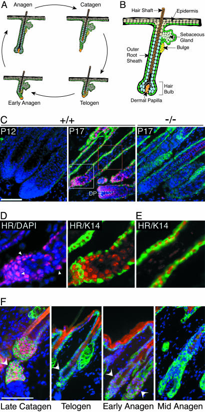
HR protein expression in hair follicles during the hair cycle. (A) Schematic representation of the hair cycle depicting changes in follicle structure. (B) Schematic of an anagen-stage hair follicle; relevant structures are indicated. A and B were adapted from ref. . (C–F) Immunofluorescent detection of HR protein (red) and keratin 14 (K14) (green). DAPI staining (blue) indicates nuclei. (C) HR protein expression in mouse back skin at P12 and P17. HR is detected in wild type (+/+) hair follicles at P17; the signal is specific because there is no staining in Hr-/- (-/-) follicles (Right). DP is denoted by white dots. Boxed areas are magnified below. (D) Magnification of region in white box shows HR staining in nuclei (HR/DAPI) and in K14-positive ORS and K14-negative hair bulb (HR/K14). Arrowheads denote nuclear staining. (E) Magnification of region in red box shows HR staining in K14-positive ORS. (F) HR expression through the hair cycle. Shown is immunofluorescent staining for HR (red) and K14 (green). Hair cycle stages are indicated. Arrowheads indicate HR signal. (Scale bars, 50 μm.)
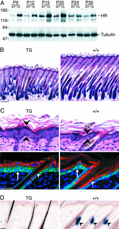
Transgenic expression of HR in progenitor keratinocytes. (A) Western analysis showing HR protein expression in wild type (WT) and K14-rHr (TG) mouse skin at the indicated postnatal ages. Detection of β-tubulin (Lower) shows protein loading. Molecular mass markers are indicated in kDa. (B) Hematoxylin–eosin staining of sections from TG and wild type (+/+) back skin at P9. (Scale bar, 100 μm). (C) (Upper) Hematoxylin–eosin staining of TG and wild type (+/+) epidermis. (Bottom) Immunofluorescent detection of K14 (green, arrows) and filaggrin (red, arrowheads). DAPI (blue) staining indicates nuclei. (D) In situ hybridization for Scd1, a sebaceous gland marker, in TG and wild type (+/+) back skin at P9. Signal is detected only in wild type (arrowheads). (Scale bar, 20 μm in C and D.)
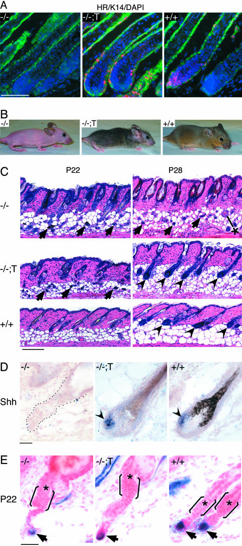
Hr expressed by the K14 promoter rescues hair regrowth in Hr-/- skin. (A) Immunofluorescent detection of HR protein (red) and K14 (green) in Hr-/- (-/-), transgenic rescue (-/-;T), and wild type (+/+) back skin. DAPI (blue) staining indicates nuclei. (Scale bar, 50 μm.) (B) Mice of the indicated genotypes at 7 weeks. (C) Hematoxylin–eosin staining of back skin sections from the indicated ages in Hr-/- (-/-), transgenic rescue (-/-;T), and wild type (+/+) mice. Arrows indicate cells in the dermis of Hr-/- and transgenic rescue mice at P22. Arrowheads indicate reformed hair bulbs in transgenic rescue and wild type mice at P28. *, cyst in Hr-/- at P28. (Scale bar, 100 μm.) (D) In situ hybridization detecting Shh mRNA in P28 mouse back skin. Shh expression (arrowheads) is detected in transgenic rescue (-/-;T) and wild type (+/+) hair follicles. Black dots outline follicle remnant in Hr-/- mice (-/-). (Scale bar, 20 μm.) (E) Alkaline phosphatase staining localizing the DP (arrows) in Hr-/- (-/-), transgenic rescue (-/-;T), and wild type (+/+) back skin at P22. Brackets approximate position of the bulge; asterisks indicate club hair. Sections are counterstained with Nuclear Fast red. (Scale bar, 20 μm.)
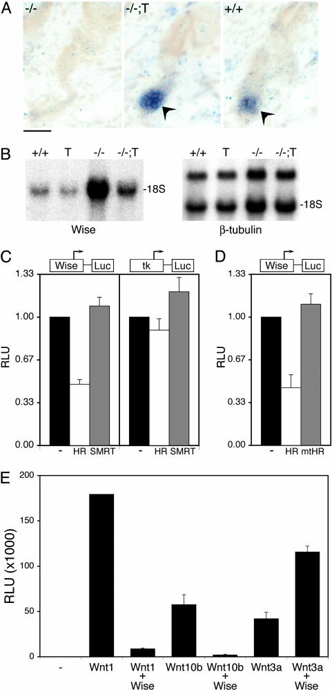
HR regulates expression of a modulator of Wnt signaling (Wise). (A) In situ hybridization for Axin2, a Wnt-responsive gene, in back skin sections from P22 mice of the indicated genotypes: -/- (Hr-/-), -/-;T (transgenic rescue), and +/+ (wild type). Arrowheads indicate Axin2 expression. (Scale bar, 20 μm.) (B) Northern analysis for Wise mRNA expression using RNA from back skin of P24 mice of the indicated genotypes: +/+ (wild type); T (K14-rHr); -/-, (Hr-/-); and -/-;T (transgenic rescue). (Right) β-tubulin hybridization of an identical blot; Wise expression was normalized to β-tubulin expression. 18S, position of 18S RNA. (C) Reporter genes for the Wise promoter (Wise-Luc) and control (tk-Luc) were transfected into cells with the indicated expression vectors. (-), empty expression vector. Results were normalized to vector control for each promoter. (D) The Wise reporter gene was transfected with expression vectors for HR or a HR derivative (mtHR) that has mutated receptor-interaction domains. Equal expression of HR and mtHR was verified by Western analysis (data not shown). (E) Cells stably expressing a Wnt-responsive reporter gene (Super TOP-Flash) were transfected with the indicated expression vectors. For B–D, relative light units (RLU) is luciferase activity relative to β-gal activity (internal control). Results are the average of at least three experiments done in duplicate.
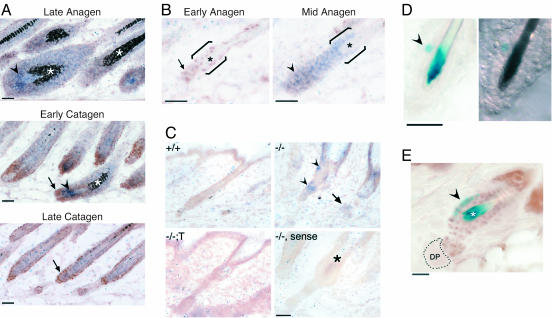
HR expression is inversely correlated with Wise mRNA expression and directly correlated with Wnt activation in the bulge. (A) In situ hybridization for Wise mRNA (blue, arrowheads) and immunohistochemistry for HR protein expression (brown, arrows) in P17 mouse back skin; follicles are at the indicated stages. Asterisks indicate pigment. (Scale bar, 50 μm.) (B) Expression of Wise mRNA (blue) and HR protein (brown) in P22 back skin; follicles are at the indicated stages. Brackets indicate bulge region; asterisks indicate club hair. (Scale bar, 50 μm.) (C) In situ hybridization for Wise mRNA in P24 mouse back skin. Expression is detected in the follicle remnant (arrowhead) and dermal cells (arrow) in Hr-/- skin (-/-). Sense control probe with adjacent Hr-/- section (-/-, sense); asterisk indicates club hair. (Scale bar, 20 μm.) (D) Detection of β-gal reporter gene expression (blue, arrowhead) in TOP-Gal mouse back skin. Bright-field (Left) and differential interference contrast (Right) microscopy images. (Scale bar, 20 μm.) (E) Detection of reporter gene expression (blue, arrowhead) and immunohistochemical staining for HR (brown). HR protein colocalizes with Wnt/β-catenin activation (blue) in the bulge. (Scale bar, 10 μm.)
References
-
- Hardy, M. H. (1992) Trends Genet. 8, 55-61. - PubMed
-
- Oshima, H., Rochat, A., Kedzia, C., Kobayashi, K. & Barrandon, Y. (2001) Cell 104, 233-245. - PubMed
-
- Cotsarelis, G., Sun, T. T. & Lavker, R. M. (1990) Cell 61, 1329-1337. - PubMed
-
- Taylor, G., Lehrer, M. S., Jensen, P. J., Sun, T. T. & Lavker, R. M. (2000) Cell 102, 451-461. - PubMed
-
- Alonso, L. & Fuchs, E. (2003) Genes Dev. 17, 1189-1200. - PubMed
Publication types
MeSH terms
Substances
Grants and funding
LinkOut - more resources
Full Text Sources
Other Literature Sources
Molecular Biology Databases
Research Materials
