. 2006 Feb 1;393(Pt 3):657-68.
doi: 10.1042/BJ20050791.

Transcriptional regulation of lysophosphatidic acid-induced interleukin-8 expression and secretion by p38 MAPK and JNK in human bronchial epithelial cells

Affiliations

Transcriptional regulation of lysophosphatidic acid-induced interleukin-8 expression and secretion by p38 MAPK and JNK in human bronchial epithelial cells

Bahman Saatian et al. Biochem J. .

Abstract

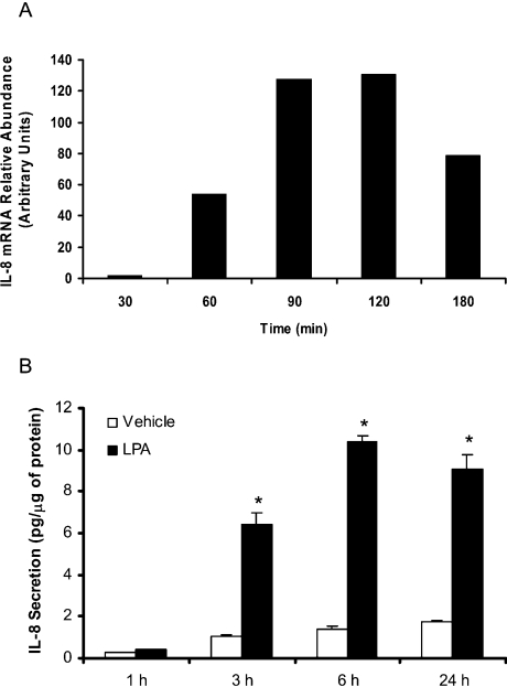
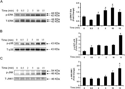
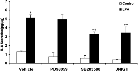
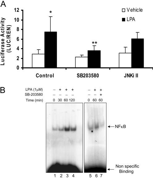
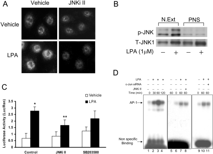
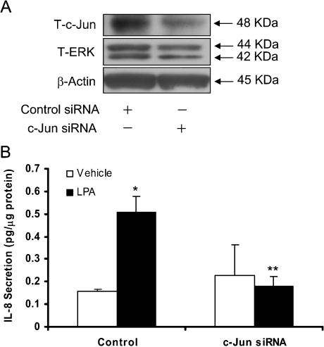
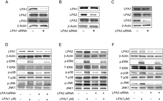
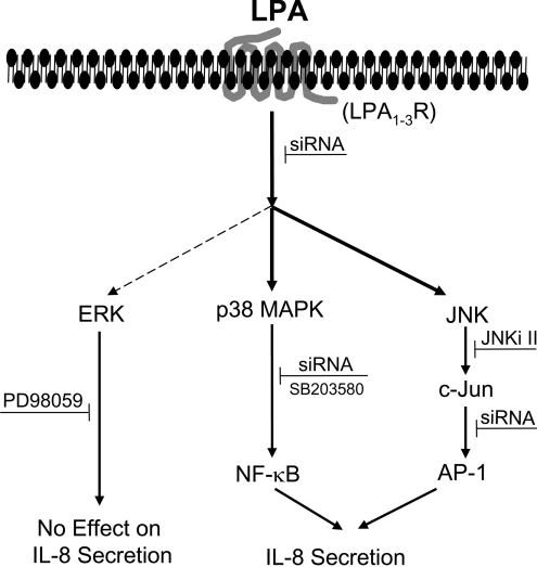
HBEpCs (human bronchial epithelial cells) contribute to airway inflammation by secreting a variety of cytokines and chemokines in response to allergens, pathogens, viruses and environmental toxins and pollutants. The potent neutrophil chemoattractant, IL-8 (interleukin-8), is a major cytokine secreted by HBEpCs. We have recently demonstrated that LPA (lysophosphatidic acid) stimulated IL-8 production in HBEpCs via protein kinase C delta dependent signal transduction. However, mechanisms of IL-8 expression and secretion are complex and involve multiple protein kinases and transcriptional factors. The present study was undertaken to investigate MAPK (mitogen-activated protein kinase) signalling in the transcriptional regulation of IL-8 expression and secretion in HBEpCs. Exposure of HBEpCs to LPA (1 microM) enhanced expression and secretion of IL-8 by 5-8-fold and stimulated threonine/tyrosine phosphorylation of ERK (extracellular-signal-regulated kinase), p38 MAPK and JNK (c-Jun N-terminal kinase). The LPA-induced secretion of IL-8 was blocked by the p38 MAPK inhibitor SB203580, by p38 MAPK siRNA (small interfering RNA), and by the JNK inhibitor JNK(i) II, but not by the MEK (MAPK/ERK kinase) inhibitor, PD98059. LPA enhanced the transcriptional activity of the IL-8 gene; that effect relied on activation of the transcriptional factors NF-kappaB (nuclear factor kappaB) and AP-1 (activator protein-1). Furthermore, SB203580 attenuated LPA-dependent phosphorylation of IkappaB (inhibitory kappaB), NF-kappaB and phospho-p38 translocation to the nucleus, NF-kappaB transcription and IL-8 promoter-mediated luciferase reporter activity, without affecting the JNK pathway and AP-1 transcription. Similarly, JNK(i) II only blocked LPA-mediated phosphorylation of JNK and c-Jun, AP-1 transcription and IL-8 promoter-mediated luciferase reporter activity, without blocking p38 MAPK-dependent NF-kappaB transcription. Additionally, siRNA for LPA(1-3) receptors partially blocked LPA-induced IL-8 production and activation of MAPKs. The LPA1 and LPA3 receptors, as compared with LPA2, were most efficient in transducing LPA-mediated IL-8 production. These results show an independent role for p38 MAPK and JNK in LPA-induced IL-8 expression and secretion via NF-kappaB and AP-1 transcription respectively in HBEpCs.


Figures

Figure 1
Figure 1. LPA stimulates IL-8 gene expression and secretion in HBEpCs
(A) HBEpCs (approx. 80% confluence in 35 mm dishes) were treated with BEGM containing 0.1% (w/v) BSA (vehicle) or BEGM containing 0.1% (w/v) BSA plus LPA (1 μM) for 30–180 min. IL-8 gene expression was evaluated by real-time RT-PCR as described in the Experimental procedures section. Results are representative of at least three independent experiments. (B) HBEpCs (approx. 80% confluence in 35 mm dishes) were treated with BEGM containing 0.1% (w/v) BSA (vehicle) or BEGM containing 0.1% (w/v) BSA plus LPA (1 μM) for 1–24 h. IL-8 in the medium was quantified by ELISA as described in the Experimental procedures section. The results are expressed as the means±S.D. (n=3) performed in triplicate. *, statistically different from control (P<0.05).
Figure 2
Figure 2. LPA stimulates phosphorylation of MAPKs in HBEpCs
HBEpCs (approx. 80% confluence in 35 mm dishes) were treated with BEGM containing 0.1% (w/v) BSA (vehicle) or BEGM containing 0.1% (w/v) BSA plus LPA (1 μM) for 0–15 min. Cell lysates (10 μg) were subjected to SDS/PAGE and Western blotted for phospho- (p) and total-(T) ERK (A) or phospho- and the total p38 MAPK (B) or phospho- and total JNK (C). Shown are representative immunoblots of three independent experiments performed in duplicate. Fold changes in phospho-ERK/ERK or phospho-p38 MAPK/p38 MAPK or phospho-JNK/JNK were calculated from the Western blots by image analysis and results were normalized to total ERK or p38 MAPK or JNK. *, significantly different from 0 min (P<0.05).
Figure 3
Figure 3. Effect of MAPK inhibitors on LPA-induced IL-8 secretion
HBEpCs (approx. 80% confluence in 35 mm dishes) were pretreated with BEGM (vehicle) or PD89059 (25 μM) or SB 203580 (25 μM) or JNKi II (25 μM) for 1 h. The cells were then challenged with BEGM containing 0.1% (w/v) BSA (vehicle) or BEGM containing 0.1% (w/v) BSA plus LPA (1 μM) for 2 h. IL-8 secreted into medium was quantified by ELISA as described in the Experimental procedures section. The results are expressed as the means±S.D. (n=3) performed in triplicate. *, significantly different from control (P<0.05), **, significantly different from cells treated with LPA alone (P<0.05).
Figure 4
Figure 4. p38 MAPK siRNA and SB-203580 attenuate LPA-induced IL-8 secretion in HBEpCs
HBEpCs (approx. 80% confluence in 35 mm dishes) were pretreated with various concentrations of SB203580 for 1 h prior to LPA (1 μM) challenge for either 15 min or 2 h. (A) Cell lysates (10 μg) from 15 min treatment were subjected to SDS/PAGE and Western blotted with phospho- and pan p38 MAPK and MAPKAPK-2 antibodies. (B) The medium was collected after 2 h LPA exposure and analysed for IL-8 by ELISA as described in the Experimental procedures section. Values are means of three independent experiments in triplicate. (C) HBEpCs (approx. 50% confluence in 35 mm dishes) were transfected with control siRNA or p38 MAPK siRNA (100 nM) for 72 h as described in the Experimental procedures section. Cell lysates (10 μg) were subjected to SDS/PAGE and Western blotted with p38 MAPK or β-actin or ERK or JNK1 antibodies. Shown is a representative blot of three independent experiments. (D) HBEpCs (approx. 50% confluence in 35 mm dishes) were transfected with control siRNA or p38 MAPK siRNA (100 nM) for 72 hours prior to LPA (1 μM) challenge for 2 h. The results are expressed as the means±S.D. (n=3) performed in triplicate. *, significantly different from control (P<0.05), **, significantly different from LPA treatment (P<0.05).
Figure 5
Figure 5. Effect of JNKi II on LPA-induced phosphorylation of JNK and c-Jun and IL-8 secretion in HBEpCs
HBEpCs (approx. 80% confluence in 35 mm dishes) were pretreated with various concentration of JNKi II for 1 h prior to LPA (1 μM) challenge for either 15 min or 2 h. (A) Cell lysates (10 μg) from 15 min treatment were subjected to SDS/PAGE and Western blotted with phosph-JNK1/2 or total JNK1 antibodies. (B) and (C) HBEpCs (approx. 80% confluence in 35 mm dishes) were pretreated with either JNKi II (25 μM) (B) or SB203580 (25 μM) (C) for 1 h prior to challenge with BEGM containing 0.1% (w/v) BSA (vehicle) or BEGM containing 0.1% BSA plus LPA (1 μM) for 15 min. Cell lysates (10 μg) were subjected to SDS/PAGE and Western blotted with phospho-c-Jun or total c-Jun antibodies. (D) Cells were exposed to LPA for 2 h and medium was collected and analysed for IL-8 by ELISA as described in the Experimental procedures section. Values are means of two independent experiments performed in triplicate.
Figure 6
Figure 6. p38 MAPK activation and NFκB transcription in HBEpCs
HBEpCs (approx. 80% confluence in 35 mm dishes) were pretreated with SB 203580 (25 μM) for 1 h prior to exposure to BEGM containing 0.1% (w/v) BSA (vehicle) or BEGM containing 0.1% (w/v) BSA plus LPA (1 μM) for 15 min. (A) Cell lysates (10 μg) were subjected to SDS/PAGE and Western blotted with phospho-IκB or phospho-p38 MAPK or phospho-MAPKAPK-2 or pan p38 MAPK or MAPKAPK-2 or β-actin antibodies. (B) as in A, cells were pretreated with JNKi II (25 μM) for 1 h prior to challenge with LPA and cell lysates (10 μg protein) were subjected to SDS/PAGE and Western blotted with phospho-IκB and β-actin antibodies. (C) and (D) HBEpCs were grown on 8-well chamber slides to approx. 70% confluence and pretreated with SB203580 (25 μM; SB) or JNKi II (25 μM) for 1 h prior to challenge with LPA (1 μM) for 15 min. Cells were then subjected to immuno-staining for NF-κB (p65) (C) or p-p38 MAPK (D) and examined by fluorescence microscopy as described in the Experimental procedures section. The immunofluorescent image is representative of two independent experiments.
Figure 7
Figure 7. Effect of MAPK inhibitors on LPA-induced NF-κB activation
A, HBEpCs (approx. 60% confluence in 24-well plates) were transiently transfected with 100 ng of NF-κB reporter construct along with 1 ng of pRL-TK reference plasmid. After overnight incubation, cells were pretreated with SB203580 (25 μM) or JNKi II (25 μM) for 1 h prior to LPA challenge (1 μM) for 3 h. Luciferase activity was measured using a commercially available kit. The results are expressed as the means±S.D. (n=3). * significantly different from vehicle controls (p < 0.05), **, significantly different from LPA treatment alone (p < 0.05). B, Nuclear extracts isolated from control (lanes1, 5) and LPA-stimulated HBEpCs from different times (lanes 2–4) were incubated with 32P end-labelled NF-κB. Lanes 6–7 contain extracts from cells pre-incubated with vehicle or SB203580 prior to LPA challenge for 60 min. Shown is a representative gel shift assay of three independent experiments.
Figure 8
Figure 8. JNKi II and c-Jun siRNA block p-JNK nuclear translocation, and LPA-induced AP-1 activation
A, HBEpCs were grown on 8-well chamber slides to approx. 70% confluence and pretreated JNKi II (25 μM) for 1 h prior to challenge with vehicle or LPA (1 μM) for 15 min. Cells were then subjected to immuno-staining with anti-phospho-JNK antibody and examined by fluorescence microscopy as described in the Experimental procedures section. The immunofluorescent image is representative of three independent experiments. (B) Nuclear extracts (N. Ext) and post-nuclear supernatant (PNS) from control (vehicle) and LPA stimulated cells (15 min) were Western blotted with anti-phospho-specific-JNK- and anti-total-JNK1- antibodies. (C) HBEpCs (approx. 60% confluence in 24-well plates) were transiently transfected with 100 ng of AP-1 reporter construct along with 1 ng of pRL-TK reference plasmid. After overnight incubation, cells pretreated with JNKi II or SB203580 for 1 h prior to LPA challenge (1 μM) for 3 h. Luciferase activity was measured using a commercially available kit. The results are expressed as the means±S.D. (n=3). * significantly different from controls (P<0.05), **, significantly different from LPA treatment (P<0.05). (D) Nuclear extracts isolated from control (lanes 1, 6, 9) and LPA-stimulated for different times (lanes 2–4) were incubated with the 32P end-labelled AP-1. Lanes 8 and 11 contain extracts from cells pretreated with JNKi II and c-Jun siRNA respectively prior to LPA challenge. Nuclear extract in lane 5 was incubated with 100-fold molar excess of unlabelled double-stranded AP-1. Shown is a representative of three independent experiments.
Figure 9
Figure 9. Effect of c-Jun siRNA on LPA-induced IL-8 secretion
HBEpCs (approx. 50% confluence in 35 mm dishes) were transfected with control siRNA or c-Jun siRNA (100 nM) for 72 h and then challenged with BEGM containing 0.1% (w/v) BSA (vehicle) or BEGM containing 0.1% (w/v) BSA plus LPA (1 μM) for 2 h. (A) Cell lysates (10 μg protein) were subjected to SDS/PAGE and Western blotted for c-Jun or ERK or β-actin. (B) Cells were treated with vehicle or LPA for 2 h and IL-8 secreted into medium was quantified by ELISA as described in the Experimental procedures section. The results are expressed as the means±S.D. (n=3) performed in triplicate. *, significantly different from controls (P<0.05), **, significantly different from LPA treatment (P<0.05).
Figure 10
Figure 10. Effect of LPA receptor siRNA on LPA-induced phosphorylation of MAPKs
HBEpCs (approx. 50% confluence in 35 mm dishes) were transfected with control siRNA or LPA1–3 receptor siRNA (200 nM) for 72 h and then harvested (A, B, C) or challenged with BEGM with 0.1% (w/v) BSA (vehicle) or BEGM with 0.1% (w/v) BSA plus LPA (1 μM) for 15 min (D, E, F). Cell lysates were subjected to SDS/PAGE and Western blotted with LPA1, LPA2 or LPA3 or β-actin antibodies (A, B, C) or anti-phospho-specific- and pan-antibodies for ERK, p38 MAPK, JNK (D, E, F).
Figure 11
Figure 11. Proposed MAPK signalling involved in LPA-induced IL-8 secretion
Ligation of LPA to its G-protein-coupled receptors LPA1–3 results in activation of ERK, p38 MAPK and JNK pathways. LPA-induced activation of p38 MAPK and JNK (heavy arrows), but not ERK (dashed arrow) regulates NF-κB and AP-1 transactivation and IL-8 secretion.

Similar articles

Cited by

References

    1. Matsukura S., Kokubu F., Noda H., Tokunaga H., Adach I. M. Expression of IL-6, IL-8, and RANTES on human bronchial epithelial cells, NCI-H292, induced by influenza virus A. J. Allergy Clin. Immunol. 1996;98:1080–1087. - PubMed
    1. Mio T., Romberger D. J., Thompson A. B., Robbins R. A., Heires A., Rennard S. I. Cigarette smoke induces interleukin-8 release from human bronchial epithelial cells. Am. J. Respir. Crit. Care Med. 1997;155:1770–1776. - PubMed
    1. Johnston S. I., Papi A., Bates P. J., Mastronarde J. G., Monick M. M., Hunninghake G. W. Low grade rhinovirus infection induces a prolonged release of IL-8 in pulmonary epithelium. J. Immunol. 1998;160:6172–6181. - PubMed
    1. Hashimoto S., Gon Y., Takeshita I., Matsumoto K., Jibiki I., Takizawa H., Kudoh S., Horie T. Diesel exhaust particles activates p38 MAP kinase to produce interleukin 8 and RANTES by human bronchial epithelial cells and N-acetylcysteine attenuates p38 MAP kinase activation. Am. J. Respir. Crit. Care Med. 2000;161:280–285. - PubMed
    1. Aggarwal A., Baker C. S., Evans T. W., Haslam P. L. G-CSF and IL-8 but not GM-CSF correlates with severity of pulmonary neutrophilia in acute respiratory distress syndrome. Eur. Respir. J. 2000;15:895–901. - PubMed

Publication types

MeSH terms