. 2005 Dec;89(6):4300-9.
doi: 10.1529/biophysj.105.061697. Epub 2005 Sep 30.

Coexistence of a two-states organization for a cell-penetrating peptide in lipid bilayer

Affiliations

Coexistence of a two-states organization for a cell-penetrating peptide in lipid bilayer

Thomas Plénat et al. Biophys J. 2005 Dec.

Abstract

Primary amphipathic cell-penetrating peptides transport cargoes across cell membranes with high efficiency and low lytic activity. These primary amphipathic peptides were previously shown to form aggregates or supramolecular structures in mixed lipid-peptide monolayers, but their behavior in lipid bilayers remains to be characterized. Using atomic force microscopy, we have examined the interactions of P(alpha), a primary amphipathic cell-penetrating peptide which remains alpha-helical whatever the environment, with dipalmitoylphosphatidylcholine (DPPC) bilayers. Addition of P(alpha) at concentrations up to 5 mol % markedly modified the supported bilayers topography. Long and thin filaments lying flat at the membrane surface coexisted with deeply embedded peptides which induced a local thinning of the bilayer. On the other hand, addition of P(alpha) only exerted very limited effects on the corresponding liposome's bilayer physical state, as estimated from differential scanning calorimetry and diphenylhexatriene fluorescence anisotropy experiments. The use of a gel-fluid phase separated supported bilayers made of a dioleoylphosphatidylcholine/dipalmitoylphosphatidylcholine mixture confirmed both the existence of long filaments, which at low peptide concentration were preferentially localized in the fluid phase domains and the membrane disorganizing effects of 5 mol % P(alpha). The simultaneous two-states organization of P(alpha), at the membrane surface and deeply embedded in the bilayer, may be involved in the transmembrane carrier function of this primary amphipathic peptide.


Figures

FIGURE 1
FIGURE 1
AFM imaging of DPPC bilayers at low P(α) concentrations. (A and C) Low-magnification height image of 0.25 (A) and 1 (C) mol % P(α) containing DPPC bilayers (top view, bar: 500 nm; z color scale: 3.0 nm); (B and D) 1 μm scan of 0.25 (B) and 1 (D) mol % P(α)-containing DPPC bilayers (3-D view, bar: 250 nm); (E) a virtual section of a pure DPPC bilayer region pierced by a hole; and (F) a virtual section of a 1 mol % P(α)-containing DPPC bilayers 500 nm scan.
FIGURE 2
FIGURE 2
AFM imaging of 2.5 mol % P(α)-containing DPPC bilayers. Low (A), intermediate (B and C), and high (D) resolution imaging of samples under PBS buffer. (A, B, and D) Top view images. (C) A 3-D view of B. Bars: 1 μm (A), 500 nm (B), and 125 nm (D).
FIGURE 3
FIGURE 3
High peptide concentrations affect bilayer formation. Topography of samples at 5 mol % (A, B, and C) and 10 mol % (D) P(α). (A) Low magnification imaging of a 5 mol % P(α)-containing DPPC bilayer sample showing the presence of adsorbed vesicles (deflection image, bar: 500 nm); (B) imaging between adsorbed vesicles (height image, bar 250 nm); (C) high magnification imaging of a 5 mol % P(α) sample (height image, bar:100 nm); and (D) low magnification imaging of a sample made with 10 mol % P(α). Note the large z color scale: 150 nm; bar: 2.5 μm.
FIGURE 4
FIGURE 4
At a low concentration, peptide filaments preferentially localize in the fluid phase of DOPC/DPPC bilayers. (A) Low magnification imaging of DOPC/DPPC (1:1) bilayer under PBS buffer (bar: 5 μm); (B) low magnification imaging of DOPC/DPPC bilayers containing 1 mol % P(α) (top view, bar: 2.5 μm); (C) 3-D view of DOPC/DPPC bilayer containing 1 mol % P(α) at intermediate magnification (bar: 1.25 μm); (D) high magnification image (top view, bar: 500 nm); and (E) a virtual section of C.
FIGURE 5
FIGURE 5
Topography of 5 mol % P(α)-containing DOPC/DPPC bilayers. (A), (B), and (D) the top view of bilayers at different magnifications (bars: 2 μm, 500 nm, and 200 nm, respectively). (C) A 3-D view of the bilayer surface at intermediate magnification. (E) A virtual section of (D).
FIGURE 6
FIGURE 6
Temperature-dependent DPH fluorescence anisotropy of P(α)-containing DPPC LUV. Solid line and solid circles correspond to the values obtained for pure DPPC. Open circles correspond to the values obtained for 1 mol % P(α) LUV. Open triangles correspond to the data obtained with 5 mol % P(α). Each individual point is the mean ± SD of three different experiments.
FIGURE 7
FIGURE 7
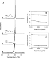
DSC heating curves of MLVs of DPPC and P(α)-DPPC mixtures. (A) DSC heating curves of (a) pure DPPC; (b) 1 mol % P(α); (c) 2.5 mol % P(α); and (d) 5 mol % P(α). (B) Effect of increasing amounts of P(α) on the peaks transition temperatures of the mixtures. (○) Main transitions. (▵) Pretransitions. (C) Effect of increasing amounts of P(α) on the transition enthalpies of P(α)-DPPC mixtures. (○) Total enthalpy. (▵) Pretransition enthalpy.

References

    1. Lindgren, M., M. Hallbrink, A. Prochiantz, and U. Langel. 2000. Cell-penetrating peptides. Trends Pharmacol. Sci. 21:99–103. - PubMed
    1. Prochiantz, A. 1996. Getting hydrophilic compounds into cells: lessons from homeopeptides. Curr. Opin. Neurobiol. 6:629–634. - PubMed
    1. Prochiantz, A. 2000. Messenger proteins: homeoproteins, TAT and others. Curr. Opin. Cell Biol. 12:400–406. - PubMed
    1. Dietz, G. P., and M. Bahr. 2004. Delivery of bioactive molecules into the cell: the Trojan horse approach. Mol. Cell. Neurosci. 27:85–131. - PubMed
    1. Drin, G., S. Cottin, E. Blanc, A. R. Rees, and J. Temsamani. 2003. Studies on the internalization mechanism of cationic cell-penetrating peptides. J. Biol. Chem. 278:31192–31201. - PubMed

Publication types

LinkOut - more resources