Retinoid X receptor ablation in adult mouse keratinocytes generates an atopic dermatitis triggered by thymic stromal lymphopoietin
- PMID: 16199515
- PMCID: PMC1253602
- DOI: 10.1073/pnas.0507385102
Retinoid X receptor ablation in adult mouse keratinocytes generates an atopic dermatitis triggered by thymic stromal lymphopoietin
Abstract
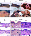
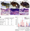
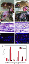
To investigate the role of retinoid X receptors (RXRs) in epidermal homeostasis, we generated RXRalphabeta(ep-/-) somatic mutants in which both RXRalpha and RXRbeta are selectively ablated in epidermal keratinocytes of adult mice. These mice develop a chronic dermatitis mimicking that observed in atopic dermatitis (AD) patients. In addition, they exhibit immunological abnormalities including elevated serum levels of IgE and IgG, associated with blood and tissue eosinophilia, indicating that keratinocyte-selective ablation of RXRs also generates a systemic syndrome similar to that found in AD patients. Furthermore, the profile of increased expression of cytokines and chemokines in skin of keratinocyte-selective RXRalphabeta-ablated mutants was typical of a T helper 2-type inflammation, known to be crucially involved in human AD pathogenesis. Finally, we demonstrate that thymic stromal lymphopoietin, whose expression is rapidly and strongly induced in RXRalphabeta-ablated keratinocytes, plays a key role in initiating the skin and systemic AD-like pathologies.
Figures


Similar articles
-
Selective ablation of Ctip2/Bcl11b in epidermal keratinocytes triggers atopic dermatitis-like skin inflammatory responses in adult mice.PLoS One. 2012;7(12):e51262. doi: 10.1371/journal.pone.0051262. Epub 2012 Dec 20. PLoS One. 2012. PMID: 23284675 Free PMC article.
-
Topical vitamin D3 and low-calcemic analogs induce thymic stromal lymphopoietin in mouse keratinocytes and trigger an atopic dermatitis.Proc Natl Acad Sci U S A. 2006 Aug 1;103(31):11736-41. doi: 10.1073/pnas.0604575103. Epub 2006 Jul 31. Proc Natl Acad Sci U S A. 2006. PMID: 16880407 Free PMC article.
-
Spontaneous atopic dermatitis in mice expressing an inducible thymic stromal lymphopoietin transgene specifically in the skin.J Exp Med. 2005 Aug 15;202(4):541-9. doi: 10.1084/jem.20041503. J Exp Med. 2005. PMID: 16103410 Free PMC article.
-
The role of thymic stromal lymphopoietin in the immunopathogenesis of atopic dermatitis.Clin Exp Allergy. 2011 Nov;41(11):1515-20. doi: 10.1111/j.1365-2222.2011.03797.x. Epub 2011 Jun 14. Clin Exp Allergy. 2011. PMID: 21672057 Review.
-
A review of the roles of keratinocyte-derived cytokines and chemokines in the pathogenesis of atopic dermatitis in humans and dogs.Vet Dermatol. 2017 Feb;28(1):16-e5. doi: 10.1111/vde.12351. Epub 2016 Jul 18. Vet Dermatol. 2017. PMID: 27426268 Review.
Cited by
-
Selective ablation of Ctip2/Bcl11b in epidermal keratinocytes triggers atopic dermatitis-like skin inflammatory responses in adult mice.PLoS One. 2012;7(12):e51262. doi: 10.1371/journal.pone.0051262. Epub 2012 Dec 20. PLoS One. 2012. PMID: 23284675 Free PMC article.
-
TRP Channels as Drug Targets to Relieve Itch.Pharmaceuticals (Basel). 2018 Oct 6;11(4):100. doi: 10.3390/ph11040100. Pharmaceuticals (Basel). 2018. PMID: 30301231 Free PMC article. Review.
-
Mediators of Chronic Pruritus in Atopic Dermatitis: Getting the Itch Out?Clin Rev Allergy Immunol. 2016 Dec;51(3):263-292. doi: 10.1007/s12016-015-8488-5. Clin Rev Allergy Immunol. 2016. PMID: 25931325 Review.
-
Thymic stromal lymphopoietin and allergic disease.J Allergy Clin Immunol. 2012 Oct;130(4):845-52. doi: 10.1016/j.jaci.2012.07.010. Epub 2012 Aug 30. J Allergy Clin Immunol. 2012. PMID: 22939755 Free PMC article. Review.
-
BP180 dysfunction triggers spontaneous skin inflammation in mice.Proc Natl Acad Sci U S A. 2018 Jun 19;115(25):6434-6439. doi: 10.1073/pnas.1721805115. Epub 2018 Jun 4. Proc Natl Acad Sci U S A. 2018. PMID: 29866844 Free PMC article.
References
Publication types
MeSH terms
Substances
LinkOut - more resources
Full Text Sources
Other Literature Sources
Molecular Biology Databases

