Comparative Study
. 2005 Oct 11;102(41):14623-8.
doi: 10.1073/pnas.0503524102. Epub 2005 Sep 30.

MAP-1 is a mitochondrial effector of Bax

Affiliations
Comparative Study

MAP-1 is a mitochondrial effector of Bax

Kuan Onn Tan et al. Proc Natl Acad Sci U S A. .

Abstract

Apoptotic stimuli induce conformational changes in Bax and trigger its translocation from cytosol to mitochondria. Upon assembling into the mitochondrial membrane, Bax initiates a death program through a series of events, culminating in the release of apoptogenic factors such as cytochrome c. Although it is known that Bax is one of the key factors for integrating multiple death signals, the mechanism by which Bax functions in mitochondria remains controversial. We have previously identified modulator of apoptosis-1 (MAP-1) as a Bax-associating protein, but its functional relationship with Bax in contributing to apoptosis regulation remains to be established. In this study, we show that MAP-1 is a critical mitochondrial effector of Bax. MAP-1 is a mitochondria-enriched protein that associates with Bax only upon apoptotic induction, which coincides with the release of cytochrome c from mitochondria. Small interfering RNAs that diminish MAP-1 levels in mammalian cell lines confer selective inhibition of Bax-mediated apoptosis. Mammalian cells with stable expression of MAP-1 small interfering RNAs are resistant to multiple apoptotic stimuli in triggering apoptotic death as well as in inducing conformation change and translocation of Bax. Similar to Bax-deficient cells, MAP-1-deficient cells exhibit aggressive anchorage-independent growth. Remarkably, recombinant Bax- or tBid-mediated release of cytochrome c from isolated mitochondria is significantly compromised in the MAP-1 knockdown cells. We propose that MAP-1 is a direct mitochondrial target of Bax.


Figures

Fig. 1.
Fig. 1.
MAP-1 is a mitochondrial protein residing at outer membrane. (A) (Left) Subcellular fractions from 293T cells were immunoblotted with the MAP-1 antibody (R5) or antibodies against the organelle-specific protein markers: COX (mitochondria), calreticulin (ER), actin (cytosol), and p14/ARF (nuclear). (Right) Sucrose gradient purified mitochondria from 293T cells were immunoblotted with R5 or anti-Cyto c antibody. (B) The N-terminal region of MAP-1 is required for mitochondrial targeting. The Myc-MAP-1 deletion mutants were transiently transfected into MCF-7 cells. Lysates from transfected cells were subjected to fractionation analysis as in A. HM and cytosolic fractions were immunoblotted with anti-myc antibody. (C) Myc-MAP-1 and the MAP-1 mutant (1–115) colocalize with mitotracker. MCF-7 cells were transiently transfected with Myc-MAP-1 or Myc-MAP-1 mutant (1–115). Sixteen hours after transfection, cells were stained with mitotracker (red) and anti-myc (green). (D) MAP-1 associates with isolated mitochondria. The indicated in vitro translated 35S-labeled proteins were incubated with mitochondria isolated from MCF-7 cells at 25°C for 20 min. Mitochondria were washed twice and repelleted by centrifugation. (E) MAP-1 is a membrane bound mitochondrial protein. Mitochondria were incubated with indicated concentration of digitonin on ice for 30 min. Mitochondria were repelleted by centrifugation (10,000 × g; 10 min) and immunoblotted for the indicated protein. (F) Mitochondrial MAP-1 is highly sensitive to prot K digestion. Mitochondria were incubated with indicated concentration of prot K on ice for 10 min. PMSF (10 mM) was added to stop prot K digestion. Mitochondria were repelleted by centrifugation and immunoblotted for the indicated protein.
Fig. 2.
Fig. 2.
Apoptotic stimuli induce association of Bax with MAP-1 and promote their integration to mitochondrial membrane. (A) Apoptotic stimuli promote endogenous Bax-MAP-1 association. MCF-7 cells were treated with STS (1 μM, 5 h) or TNF (40 ng/ml, 7 h). Cell lysates were immunoprecipitated with anti-Bax antibody. Immunoprecipitates were immunoblotted with MAP-1 (M6) or Hsp60 antibody (Upper). Total lysates were immunoblotted with the indicated antibodies (Lower). (B) Apoptotic stimuli promote integration of both MAP-1 and Bax into mitochondrial outer membrane. Mitochondria isolated from control or TNF-treated (40 ng/ml, 7 h) MCF-7 cells were resuspended in 0.1M Na2CU3 (pH 11.5), where indicated, and incubated on ice for 20 min followed by sonication for 5 min. Mitochondria were repelleted by centrifugation (350,000 × g, 20 min) and immunoblotted for the indicated protein.
Fig. 3.
Fig. 3.
MAP-1 is required for Bax-induced, but not Bak-induced, apoptosis. (A) MCF-7 cells were first transfected with scramble siRNA (Scr) or MAP-1 siRNAs R1 or R3 for 2 days followed by a second transfection with pXJ40 vector, HA-Bax, or HA-Bak together with the GFP reporter. Apoptosis was determined by counting the percentage of GFP-positive cells that exhibited condensed nuclei morphology. (B)(Left) HCT116 Bax+/- or Bax-/- cells were subjected to identical transfection protocols as in A, and the cells were processed 18 h after the second transfection. (Right) MAP-1 induces apoptosis in Bax+/- and Bax-/- cells with similar efficacy. HCT116 Bax+/- or Bax-/- cells were transiently transfected with either pXJ40 vector, HA-Bax, or Myc-MAP-1. The percentage apoptosis was determined as in A. Results are presented as percentage of control (mean ± SD, n = 3).
Fig. 4.
Fig. 4.
MAP-1 knockdown MCF-7 cells are resistant to diverse apoptotic stimuli. (A) MAP-1 protein level is substantially reduced in SM-R3-12 cells stably expressing the MAP-1 siRNA. Cell lysates were immunoprecipitated with Bax (N20) or MAP-1 (R5) antibodies followed by immunoblotting with MAP-1 (M6) or Bax (2D2) antibodies. (B) MAP-1 knockdown cells are resistant to apoptotic death triggered by STS. (Top) Cells were harvested and stained with JC-1 for analysis of mitochondrial membrane potential change by flow cytometry. (Middle) The extent of caspase activation following treatment with STS was assayed with AC-DEVD-AFC. (Bottom) Cell viability after STS treatment was determined by WST-1 assay. (CE) MAP-1 knockdown cells displayed resistance to apoptosis triggered by TNF (C), UV (D), and serum withdrawal (E). Cells were subjected to various apoptotic insults as indicated and the dose-dependent cell viability responses were determined by the following assays: JC-1 (TNF) or trypan blue exclusion (UV and serum withdrawal). Results are presented as percentage of control (mean ± SD, n = 3).
Fig. 5.
Fig. 5.
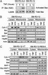
Apoptotic stimuli-mediated conformation change and translocation of Bax are inhibited in MAP-1-depleted cells. (A) TNF-mediated conformation change of Bax was affected in MAP-1 knockdown cells. Equal amounts of total lysates from the TNF-treated (40 ng/ml) cells were immunoprecipitated with the conformation-specific anti-Bax antibody, N20. Cell lysates and immunoprecipitates were immunoblotted with anti Bax. (B) MAP-1 is required for apoptosis-mediated Bax translocation and Cyto c release. Cytosolic and mitochondrial fractions from cells treated with either STS (1 μM, 5 h) or TNF (40 ng/ml,10 h) were immunoblotted with the indicated antibodies. (C) Stable expression of myc-MAP-1 in the MAP-1 knockdown cells is sufficient for restoring the sensitivity of MAP-1-deficient cells to apoptotic stimuli-mediated Bax translocation and Cyto c release. Cells were treated and analyzed as in B.
Fig. 6.
Fig. 6.
MAP-1 is required to facilitate Bax- and tBid-mediated release of Cyto c from isolated mitochondria. (A) Mitochondria isolated from control or MAP-1 knockdown cells were incubated with recombinant Bax, tBid, or Bak followed by centrifugation. The supernatants and pellets were immunoblotted with indicated antibodies. (B) Stable expression of myc-MAP-1 in MAP-1 knockdown cells restores the sensitivity of mitochondria to Bax- and tBid-induced release of Cyto c. Mitochondria isolated from MAP-1 knockdown or rescue cells were treated and analyzed as in A.(C) In vitro translated MAP-1 restores the sensitivity of MAP-1-deficient mitochondria to Bax-mediated release of Cyto c. Mitochondria from MAP-1 knockdown cells were preincubated with rabbit reticulate lysate (R. lysates), 35S-labeled in vitro translated MAP-1, or VDAC (1 × 10⁵ cpm) for 20 min at 25°C. The mitochondria was washed twice, treated with recombinant Bax, and analyzed as in A.

References

    1. Wang, X. (2001) Genes Dev. 15, 2922-2933. - PubMed
    1. Green, D. R. & Reed, J. C. (1998) Science 281, 1309-1312. - PubMed
    1. Cory, S. & Adams, J. M. (2002) Nat. Rev. Cancer 2, 647-656. - PubMed
    1. Danial, N. N. & Korsmeyer, S. J. (2004) Cell 116, 205-219. - PubMed
    1. Cartron, P. F., Gallenne, T., Bougras, G., Gautier, F., Manero, F., Vusio, P., Meflah, K., Vallette, F. M. & Juin, P. (2004) Mol. Cell 16, 807-818. - PubMed

Publication types