. 2005 Dec 2;280(48):40201-9.
doi: 10.1074/jbc.M505186200. Epub 2005 Sep 30.

Identification of a novel extracellular cation-sensing G-protein-coupled receptor

Affiliations

Identification of a novel extracellular cation-sensing G-protein-coupled receptor

Min Pi et al. J Biol Chem. .

Abstract

The C family G-protein-coupled receptors contain members that sense amino acid and extracellular cations, of which calcium-sensing receptor (CASR) is the prototypic extracellular calcium-sensing receptor. Some cells, such as osteoblasts in bone, retain responsiveness to extracellular calcium in CASR-deficient mice, consistent with the existence of another calcium-sensing receptor. We examined the calcium-sensing properties of GPRC6A, a newly identified member of this family. Alignment of GPRC6A with CASR revealed conservation of both calcium and calcimimetic binding sites. In addition, calcium, magnesium, strontium, aluminum, gadolinium, and the calcimimetic NPS 568 resulted in a dose-dependent stimulation of GPRC6A overexpressed in human embryonic kidney cells 293 cells. Also, osteocalcin, a calcium-binding protein highly expressed in bone, dose-dependently stimulated GPRC6A activity in the presence of calcium but inhibited the calcium-dependent activation of CASR. Coexpression of beta-arrestins 1 and 2, regulators of G-protein signaling RGS2 or RGS4, the RhoA inhibitor C3 toxin, the dominant negative Galpha(q)-(305-359) minigene, and pretreatment with pertussis toxin inhibited activation of GPRC6A by extracellular cations. Reverse transcription-PCR analyses showed that mouse GPRC6A is widely expressed in mouse tissues, including bone, calvaria, and the osteoblastic cell line MC3T3-E1. These data suggest that in addition to sensing amino acids, GPRC6A is a cation-, calcimimetic-, and osteocalcin-sensing receptor and a candidate for mediating extracellular calcium-sensing responses in osteoblasts and possibly other tissues.


Figures

FIGURE 1
FIGURE 1. Comparison of conserved calcium and calcimimetic binding residues in members of the family C GPCRs.
A, extracellular region containing calcium binding sites. The amino acids required for calcium sensing in CASR, Ser-147, Ser-169, and Pro-823 (boxed residues) but not Ser-170 (*) are conserved between CASR and GPRC6A. m-, mouse; h-, human. B, a segment of the transmem-brane region containing putative calcimimetic binding sites. The Glu-837 binding site for calcimimetics in CASR is conserved in GPRC6A but not mGluRs. Regions of the extracellular domain and transmembrane domain from human and mouse GPRC6A (NM_148963 for human GPRC6A, and NM_153071 for mouse GPRC6A) were aligned with the human CASR (NM_000388) amino acid sequence and with members of the mGluRs (NM-000838 for human mGluR1, NM_000840 for human mGluR3, and NM_000842 for human mGluR5) with calcium-sensing properties.
FIGURE 2
FIGURE 2. Cell surface expression of GRPC6A.
A, confocal microscopy images of HEK-293 cells transiently transfected with the Myc-tagged mouse GPRC6A. B, non-transfect HEK-293 controls. GPRC6A was detected with an anti-c-Myc fluorescein isothiocyanate antibody. Left panel, representative view using light microscopy (LM). Right panel, the same view under fluorescent microscopy (FL), magnification 100×. Immunofluorescence was observed in a peripheral pattern consistent with cell surface membrane expression.
FIGURE 3
FIGURE 3. Effect of various extracellular cations on GPRC6A-mediated EKR activation.
A, comparison of dose-dependent effects of Ca2+on GPRC6A- and CASR-mediated ERK activation. Extracellular calcium activated both receptors, but GPRC6A responded to higher concentrations of extracellular calcium. B, cation specificity of GPRC6A activation. In addition to Ca2+, Gd3+ (100 μm) and Sr2+ (40 mm) as well as Mg2+ and Al3+ stimulated GPRC6A-mediated ERK activation. Magnesium and aluminum, similar to calcium (A) also resulted in a dose-dependent stimulation of ERK activity in HEK-293 cells transfected with GPRC6A. ERK was not activated by cations in controls HEK-293 cells without GPRC6A. C, the calcimimetic NPS R568 stimulates GPRC6A-mediated EKR activity. Application of the active isomer NPS R568 resulted in a dose-dependent stimulation of GPRC6A-mediated ERK activation (at concentrations of 0.5 μm) in the presence of 1 m m extracellular Ca2+, but the less active isomer NPS S568 was effective only at concentrations 5 μm. The HEK-293 cells transfected with GRPC6A or CASR were incubated in Dulbecco’s modified Eagle’s medium/F-12 containing 0.1% bovine serum albumin quiescence media and exposed to the various cations or calcimimetics for 10 min, and ERK activation was determined as described under “Experimental Procedures.”
FIGURE 4
FIGURE 4. Effects of polyvalent cations on GPRC6A-induced SRE-reporter gene activity and intercellular calcium in HEK-293 cells.
A, dose-dependent effects of various cations, Ca2+, Mg2+, Sr2+, and Gd3+, on SRE-luciferase activity in HEK-293 cells stably transfected with human GPRC6A cDNA and the SRE-luciferase reporter construct. Growth arrest was induced in 3-day-old subconfluent cultures by preincubation for 24 h under serum-free conditions before the addition of cations at the indicated concentrations. Luciferase activities were measured as described under “Experimental Procedures.” The values depicted represent the mean ± S.E. B, GPRC6A-mediated intracellular calcium mobilization in response to Ca2+ and Sr2+. HEK-293/Gqi5 cells stably expressing GPRC6A were seeded at 2 × 10⁴ cells/well and loaded with Calcium 3 dye (Molecular Probes). Cells were treated with 5, 40, and 80 mm Ca2+ or Sr2+, and fluorescence was measured using Fluorescence Imaging Plate Reader as described under “Experimental Procedures.” The graph shows the magnitude of the response due to GPRC6A. RFU, relative fluorescence units.
FIGURE 5
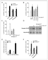
FIGURE 5. G-protein coupling of GPRC6A.
A, overexpression of β-arrestins block GPRC6A-mediated activation of the SRE. HEK-293 cells were cotransfected SRE-luciferase and pCMV-β-gal (0.015 μg) along with the construct (1.0 μg) directing the expression of β-arrestin 1 or β-arrestin 2. B, inhibition of GPRC6A activity by expression of RGS2, -4, -12. HEK-293 cells were cotransfected with expression vectors for RGS2, RGS4, or RGS12 (1 μg) along with GPRC6A (0.5 μg), the SRE-luciferase reporter gene (0.01 μg), and pCMV-β-gal (0.015 μg). C and D, pertussis toxin (PT) inhibits GPRC6A activation of the SRE (C) and ERK (D). HEK-293 cells, which stably cotransfected GPRC6A and SRE-luciferase plasmid DNAs, were cultured in serum-free media for 24 h. After pretreatment with 100 ng/ml pertussis toxin for 5 h, the cells were stimulated by 10 mm calcium. E, effect of activated mutants of Gαq subunits on the activity of the SRE. HEK-293 cells were cotransfected with 0.5 μg of expression vectors for the constitutively activated mutant of Gαq (Gαq QL) along with expression vectors for GPRC6A (0.5 μg) or empty vector, the SRE-luciferase reporter gene (0.01 μg), and p-cytomegalovirus-β-galactosidase (pCMV-β-gal) (0.015 μg). F, GPRC6A activity is inhibited by the expression of Gαq minigene construct, Gαq-(305–359), and Rho A-specific inhibitor C3 toxin. HEK-293 cells were cotransfected with the constructs directing the expression of Gαq-(305–359) (1.0 μg) or C3 toxin construct (1.0 μg) with the GPRC6A (0.5 μg), the SRE-luciferase reporter gene (0.01 μg), and pCMV-β-gal (0.015 μg). Data are shown as relative luciferase activity reported as the percent induction compared with the activity under non-stimulated conditions and normalized for β-galactosidase. Values represent the mean ± S.E. of at least three experiments. Values sharing the same superscript are not significantly different at p < 0.05.
FIGURE 6
FIGURE 6. Expression of GPRC6A in mouse tissues and osteoblasts.
A, expression pattern of GPRC6A from the mouse cDNA panel. PCR products were amplified from multiple tissue cDNA panels that contain normalized adult mouse cDNA preparations. kb, kilobase. B, GPRC6A expression in mouse tissues by RT-PCR. The primers for GPRC6A application are GPRC6A.F737 (tgtgcattgccttcaaagag) and GPRC6A.R1812 (gagagccaaggagtcatccc). C, GPRC6A expression in mouse bone tissues and osteoblastic cell line MC3T3-E1 by RT-PCR. The primers for GPRC6A application are GPRC6A.F1321 (gctcgagactgcaagaaacc) and GPRC6A.R2320 (tgaaggccagaactgtgatg). −RT indicates negative control from omitting the reverse transcription step. We used the housekeeping control gene glyceraldehyde-3-phosphate dehydrogenase (G3PDH) for a positive control of RNA integrity.
FIGURE 7
FIGURE 7. Osteocalcin is a co-factor for GPRC6A.
A, dose-dependent enhancement by osteocalcin of calcium-mediated activation of GPRC6A. B, osteocalcin augments strontium-and magnesium-stimulated SRE-luciferase activation by GPRC6A transfected into HEK-293 cells but not the effect of aluminum. C, comparison of osteocalcin activation of CASR. Ob. CASR and GPRC6A. HEK-293 cells transfected with GPRC6A respond to osteocalcin in the presence of extracellular calcium similar to MC3T3-E1 osteoblasts. Osteocalcin inhibits calcium stimulation of CASR transfected into HEK-293 cells. Values represent the mean ± S.E. of at least three experiments. Values sharing the same superscript are not significantly different at p < 0.05. con, control.

Similar articles

Cited by

References

    1. Hofer AM, Brown EM. Nat Rev Mol Cell Biol. 2003;4:530–538. - PubMed
    1. Chattopadhyay N, Brown EM. Cell Signal. 2000;12:361–366. - PubMed
    1. VanHouten J, Dann P, McGeoch G, Brown EM, Krapcho K, Neville M, Wysolmerski JJ. J Clin Investig. 2004;113:598–608. - PMC - PubMed
    1. Cheng SX, Okuda M, Hall AE, Geibel JP, Hebert SC. Am J Physiol Gastrointest Liver Physiol. 2002;283:240–250. - PubMed
    1. Conigrave AD, Quinn SJ, Brown EM. Proc Natl Acad Sci U S A. 2000;97:4814–4819. - PMC - PubMed

Publication types

MeSH terms